Ultrasonography has become a fundamental diagnostic tool in equine reproductive medicine, offering veterinarians a real-time, non-invasive, and highly informative method to assess the uterine health of mares, especially those presenting with infertility. Across equine breeding practices in Europe, North America, and Australia, real-time ultrasound is increasingly relied upon to diagnose uterine pathologies that were once only detectable through biopsy, palpation, or labor-intensive laboratory work. This article explores how equine practitioners utilize ultrasound to evaluate uterine abnormalities in infertile mares, outlining a clinical approach that merges global best practices with practical insight.

The Importance of Uterine Health in Mare Fertility
Reproductive success in mares largely depends on the condition of the uterus, which must support early embryonic development, placental attachment, and fetal growth. Even minor uterine abnormalities can hinder this process, leading to early embryonic loss or failure to conceive altogether. In infertile mares—whether barren, repeat breeders, or those with a history of embryonic loss—evaluating uterine health is a critical first step in identifying the underlying cause.
Internationally, veterinarians have emphasized the role of uterine infections (endometritis), fluid accumulation (intrauterine fluid or "IUF"), cysts, and fibrosis as the most common pathological findings contributing to subfertility. Fortunately, real-time ultrasound allows for efficient and repeatable detection of these abnormalities.
The Clinical Utility of Real-Time Ultrasound
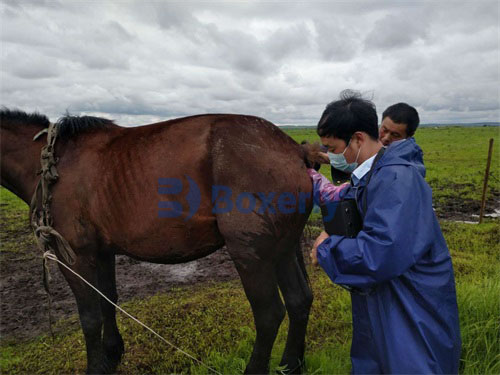
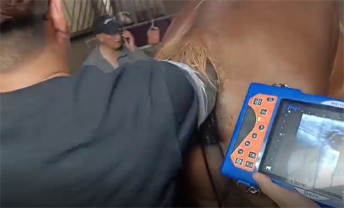
Modern real-time ultrasound scanners produce high-resolution B-mode images that reveal the internal structures of the uterus in real time. Transrectal ultrasonography is the most widely used technique due to its convenience, safety, and accessibility. In countries like the USA and the UK, this method is considered the gold standard for reproductive evaluation.
Veterinarians perform ultrasonography during estrus, diestrus, and post-breeding to capture the uterus under different hormonal influences. Key features evaluated include:
Uterine wall thickness and uniformity
Presence and echogenicity of intrauterine fluid
Endometrial cysts
Fibrosis or scar tissue
Uterine tone and contractility
Post-breeding inflammatory response
Each of these findings contributes valuable information toward making a diagnosis and formulating a treatment plan.
Identifying Intrauterine Fluid Accumulation
One of the most common findings in infertile mares is intrauterine fluid accumulation. On ultrasound, this appears as an anechoic (black) or hypoechoic (dark grey) area within the uterine lumen. In many cases, IUF is associated with poor uterine clearance mechanisms or post-breeding endometritis.
Veterinarians in Australia and Canada routinely use ultrasound to monitor how quickly fluid is cleared post-breeding. Mares that fail to clear fluid within 12 to 24 hours may require oxytocin therapy, uterine lavage, or antimicrobial treatment.
Ultrasound-guided observation of uterine fluid also enables veterinarians to distinguish between physiological fluid (such as that seen in estrus) and pathological fluid due to inflammation or infection. This is critical for determining whether a mare is suitable for immediate breeding or requires intervention.
Diagnosis of Endometrial Cysts
Endometrial cysts are another frequent finding, particularly in older or multiparous mares. They appear as small, round to oval, anechoic to hyperechoic structures embedded in the uterine lining. While not all cysts cause infertility, large or numerous cysts can impede embryo migration—a necessary step for maternal recognition of pregnancy.
Real-time ultrasound allows precise mapping of cyst location and size. Practitioners in the UK often create cyst charts during breeding season to differentiate these benign structures from early embryonic vesicles. This practice minimizes false-positive pregnancy diagnoses and improves reproductive decision-making.
Assessing Uterine Fibrosis (Endometrosis)
Uterine fibrosis, also called endometrosis, involves the formation of fibrotic tissue within the endometrium, reducing its ability to support pregnancy. Unlike cysts or fluid, fibrosis can be more subtle on ultrasound.
Using a high-frequency linear probe, veterinarians in Germany and the Netherlands have reported improved sensitivity in detecting endometrial heterogeneity—a hallmark of fibrotic change. This heterogeneity appears as irregular echo textures or echogenic streaks within the uterine lining.
Although endometrial biopsy remains the gold standard for definitive diagnosis of fibrosis, ultrasound is an invaluable screening tool and helps guide whether more invasive diagnostics are warranted.

The Post-Breeding Inflammatory Response
After breeding, all mares experience a mild inflammatory response, but susceptible individuals fail to resolve this inflammation, leading to persistent breeding-induced endometritis (PBIE). Through sequential ultrasound exams—typically at 6, 12, and 24 hours post-breeding—veterinarians in the U.S. and Argentina monitor for retained fluid and abnormal endometrial echotexture, both signs of PBIE.
Early identification through ultrasound allows immediate therapeutic intervention, including:
Uterine lavage
Oxytocin or prostaglandin administration
Intrauterine antibiotics or antiseptics
Mares that respond poorly may benefit from breeding with frozen semen via deep horn insemination, which reduces uterine contamination.
Equipment and Probe Selection
Veterinary practitioners emphasize the importance of using appropriate ultrasound equipment. Linear and convex probes ranging from 5 to 7.5 MHz are commonly used, providing an optimal balance between penetration and image resolution.
In field settings, especially on large breeding farms in the U.S. and Australia, portable ultrasound systems such as the BXL-V50 have gained popularity. This model offers:
High-definition real-time imaging
Transrectal and abdominal compatibility
Durable, waterproof casing ideal for rugged environments
Long battery life—ideal for examining multiple mares in succession
Such devices enhance the consistency and accuracy of reproductive monitoring, regardless of field conditions.
Integrating Ultrasound with Breeding Strategies
Veterinarians and breeders increasingly rely on ultrasound to tailor breeding strategies for each mare. By integrating ultrasonographic findings with hormone profiles (e.g., progesterone and estrogen levels), practitioners can:
Time insemination accurately
Identify silent heats or ovulation failure
Decide whether to delay breeding due to uterine pathology
Track treatment success (e.g., resolution of fluid after therapy)
In countries like France and New Zealand, this holistic approach has become standard, reducing both breeding costs and time to conception.
The Role of Ultrasound in Repeat Breeder Mares
Repeat breeder mares—those that fail to conceive after multiple cycles—require intensive evaluation. In these cases, ultrasound can reveal subtle uterine abnormalities that were missed in previous cycles. Practitioners commonly evaluate:
Uterine horn symmetry and size
Presence of endometrial edema during estrus
Echogenic changes in response to hormonal stimulation
Dynamic changes post-treatment (e.g., uterine flushing, antibiotics)
These insights allow clinicians to adjust protocols and improve outcomes, especially when combined with embryo transfer or advanced reproductive techniques.

Limitations and Future Prospects
While ultrasonography is powerful, it has limitations. Some abnormalities, such as deep fibrosis or microscopic inflammation, may go undetected. Additionally, operator experience and probe quality influence diagnostic accuracy.
However, emerging technologies—including contrast-enhanced ultrasound, elastography (measuring tissue stiffness), and 3D imaging—hold promise for enhancing diagnostic detail. In research settings, such as those at Colorado State University and the University of Guelph, these tools are already showing improved sensitivity in detecting endometrial pathology.
Conclusion
Real-time ultrasound has revolutionized the diagnosis and management of uterine abnormalities in infertile mares. From detecting fluid and cysts to assessing uterine tone and post-breeding response, ultrasound provides a dynamic, evidence-based view into the reproductive health of the mare.
By following a structured, clinical approach, veterinarians can accurately diagnose and monitor conditions that compromise fertility. When paired with timely treatment and individualized breeding strategies, this technology greatly improves the reproductive success rate of mares across the globe.
As portable ultrasound systems like the BXL-V50 continue to evolve, equine practitioners are better equipped than ever to address infertility in mares with confidence, efficiency, and precision.
References
Samper, J. C., & Pycock, J. (2021). Equine Breeding Management and Artificial Insemination, 3rd ed. Elsevier.
Watson, E. D., & Barbacini, S. (2020). “Ultrasonographic Assessment of Equine Uterine Pathology: A Clinical Guide.” Veterinary Clinics of North America: Equine Practice, 36(1), 1–22.
Brinsko, S. P., et al. (2011). Manual of Equine Reproduction, 3rd ed. Mosby.
Renaudin, C. D. (2023). “Real-Time Ultrasound in Equine Reproduction.” TheHorse.com.
McKinnon, A. O., & Squires, E. L. (2011). Equine Reproduction, 2nd ed. Wiley-Blackwell.


