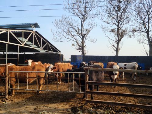
Performance horses—whether used in racing, jumping, dressage, or eventing—depend on the health and integrity of their tendons for optimal athletic function. Tendon injuries not only impair performance but also threaten a horse's long-term soundness and career longevity. Among various diagnostic tools, ultrasound imaging has become a gold standard for evaluating tendon lesions in equine athletes. In countries like the United States, United Kingdom, Germany, and Australia, where equestrian sports are both culturally embedded and economically significant, veterinarians rely heavily on ultrasound to detect, monitor, and manage tendon injuries effectively.
")
This article explores the diagnostic value of ultrasound imaging in detecting tendon lesions of the performance horse, highlighting its importance, methodology, clinical applications, limitations, and future developments. Drawing on current international practices, scientific literature, and veterinary field experience, we aim to provide a comprehensive, readable, and evidence-based discussion for horse owners, trainers, and veterinary professionals alike.
Understanding Tendon Injuries in Performance Horses
Tendon injuries are among the most common causes of lameness in horses engaged in strenuous physical activity. The superficial digital flexor tendon (SDFT) and deep digital flexor tendon (DDFT) are particularly vulnerable due to the intense repetitive strain they endure during exercise. Injuries range from mild fiber disruptions to complete tendon rupture.
Common causes include:
Repetitive microtrauma from overuse
Sudden overstretching (e.g., during jumping or missteps)
Poor conformation or hoof imbalance
Training errors or inadequate warm-ups
Tendon injuries are often insidious. Clinical signs like swelling, heat, or lameness may appear only after significant damage has occurred. Early detection is essential to prevent irreversible changes such as fibrosis or re-injury after recovery.
Why Ultrasound Imaging?
In countries with advanced equine sports industries—such as the UK (home to Ascot), the USA (with its thriving Thoroughbred racing), and Australia (where equestrian sports are Olympic mainstays)—ultrasound imaging is universally considered the first-line modality for diagnosing and monitoring tendon injuries. Unlike X-rays, which are ideal for bones, ultrasound excels in soft tissue visualization, offering detailed, real-time imaging of tendon fibers, sheaths, and surrounding structures.
Benefits of Ultrasound in Tendon Evaluation:
Non-invasive and safe: No radiation exposure; suitable for repeated use
Portable and real-time: Can be used stalls-side or at competition grounds
Sensitive to subtle changes: Detects fiber disruptions, edema, and neovascularization
Quantitative tracking: Enables measurement of lesion size and echogenicity over time
Guidance for treatment: Assists in therapeutic procedures such as stem cell or PRP injections
Ultrasound Technique and Protocols in Equine Tendon Imaging
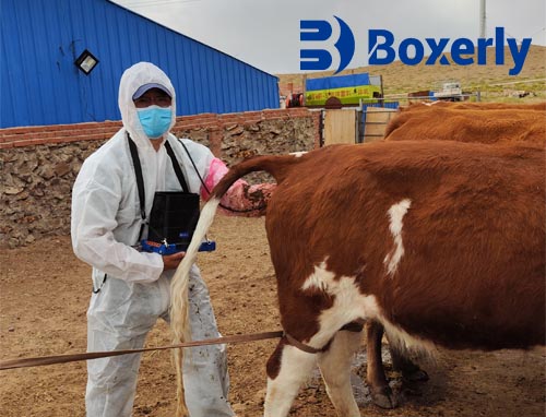
Ultrasound imaging in performance horses is typically performed using a 7.5 to 12 MHz linear probe, which offers high-resolution images for superficial structures like tendons. The horse should be restrained in a quiet environment, and the limb cleaned, clipped, and applied with coupling gel for optimal image quality.
Imaging Planes:
Longitudinal plane (sagittal): Assesses fiber alignment and longitudinal tears
Transverse plane (axial): Measures tendon cross-section and identifies focal lesions
Color Doppler mode: Detects abnormal blood flow associated with healing or inflammation
A standardized 12-zone approach (dividing the metacarpal/metatarsal region into equal sections) is often used internationally for consistent lesion tracking and reporting.
What Veterinarians Look For
Key indicators of tendon lesions on ultrasound include:
Hypoechoic (dark) areas: Represent fluid or fiber disruption
Increased cross-sectional area (CSA): Indicates swelling and inflammation
Loss of fiber pattern: Suggests partial or complete tearing
Peritendinous effusion: Suggests tenosynovitis or sheath injury
Neovascularization (seen on Doppler): Indicates active healing or chronic pathology
Veterinarians in the UK and US often rely on grading systems like the Echogenicity Score or Lesion Percentage System to quantify injury severity and monitor progression objectively.
")
Monitoring Healing and Guiding Rehabilitation
Ultrasound imaging is not just diagnostic—it is pivotal in tracking tendon healing. Periodic scans every 30–45 days allow veterinarians to evaluate:
Reduction in lesion size
Restoration of fiber alignment
Normalization of cross-sectional area
This evidence-based monitoring helps tailor rehabilitation plans. In the US, equine rehab centers increasingly integrate ultrasound in their exercise progression protocols—deciding when to transition from hand-walking to trotting, or to reintroduce jumping—based on tendon healing observed on scan.
Real-Life Case Example
Dr. Sue Dyson, a renowned British equine lameness specialist, reported in her 2023 lecture series how a 7-year-old eventing gelding presented with slight swelling and no overt lameness. While palpation was inconclusive, ultrasound detected a 15% fiber disruption in the SDFT in Zone 3C. Immediate rest and biologic therapy followed by structured rehab helped the horse return to full competition six months later—a success only made possible by early ultrasonographic detection.
International Research on Ultrasound's Diagnostic Power
Numerous peer-reviewed studies reinforce ultrasound’s clinical relevance:
Rantanen et al. (2003) showed that ultrasonography detected 85% of early-stage SDFT lesions missed by palpation alone.
Smith et al. (2022, Equine Veterinary Education) confirmed that ultrasound outperformed thermography and palpation in detecting subclinical tendonitis in jumpers.
Australian Equine Diagnostic Institute (2021) published findings on how serial ultrasound predicted recurrence risk with 92% accuracy—an insight valuable for training schedules.
Challenges and Limitations
Despite its many advantages, ultrasound is not without limitations:
Operator dependence: Image quality and interpretation vary by practitioner skill
Limited penetration: Deep structures (e.g., suspensory branches or proximal tendons) can be difficult to image in large horses
Cannot determine mechanical strength: Even visually healed tendons may be biomechanically weak
May miss tiny microtears: Particularly during the very early phase before edema forms
To overcome these, some practitioners combine ultrasound with MRI or thermography in complex cases, especially in high-value sport horses.
Future Trends and Technology
The field of equine ultrasonography is evolving rapidly. portable ultrasound machines like the BXL-V50 now offer high-definition imaging, longer battery life, and real-time Doppler capabilities—all crucial for field use. These features make it easier for vets to perform tendon exams at racecourses, competitions, or breeding farms.
In the future, AI-assisted image interpretation and 3D tendon reconstruction may become commonplace. International institutions, including the University of Florida College of Veterinary Medicine, are already researching algorithms to automate lesion detection and improve diagnostic consistency.
Conclusion
Ultrasound imaging has revolutionized the way veterinarians diagnose, monitor, and manage tendon injuries in performance horses. With its non-invasive nature, real-time capability, and precise visualization of soft tissue structures, it serves as a critical tool in equine sports medicine.
Whether you're a trainer in Kentucky, a veterinarian in Bavaria, or a rider in Sydney, understanding how and why to use ultrasound could be the difference between a short break and a career-ending injury. As technology improves and awareness spreads, the use of ultrasound in tendon care is poised to grow, enhancing horse welfare and extending equine athletic careers worldwide.
References
Rantanen, N. W., & Genovese, R. L. (2003). Tendon and Ligament Injury. In Diagnosis and Management of Lameness in the Horse. Saunders.
Smith, L., Browne, H., & Dyson, S. (2022). Ultrasound imaging as a predictor of recurrence risk in equine superficial digital flexor tendonitis. Equine Veterinary Education, 34(3), 147–155.
Australian Equine Diagnostic Institute. (2021). “Ultrasound Use in High-Performance Horses.”
Whitaker, D. A., & Smith, E. (2021). Veterinary Ultrasonography in Food-Producing Animals. Journal of Veterinary Imaging.
University of Florida College of Veterinary Medicine. (2023). “AI and Ultrasound for Equine Tendon Injury Detection.”