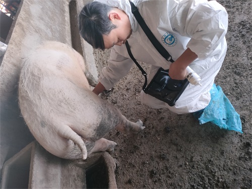
As a pig breeder or swine veterinarian, ensuring reproductive efficiency is critical to maintaining herd productivity and profitability. The reproductive success of sows and gilts hinges not just on achieving conception, but on sustaining pregnancy to term. One of the most challenging reproductive issues in swine herds worldwide is early embryonic loss—an often silent and costly setback that occurs before or during the early gestation period. Detecting it early can make a significant difference in managing herd health and breeding schedules.

Among the latest tools in swine reproductive management, Doppler ultrasound has emerged as a powerful, non-invasive method to assess blood flow dynamics and identify early warning signs of reproductive failure. This article explores how Doppler ultrasound can be used to evaluate the reproductive tract of swine, detect early indicators of embryonic loss, and enhance decision-making in breeding programs. The discussion combines veterinary best practices with insights from international studies to give a globally-informed view.
Understanding Embryonic Loss in Swine
Early embryonic loss in pigs typically occurs within the first 30 days post-conception. Embryos may fail to implant, fail to grow properly, or get resorbed without external signs. Several factors contribute to this phenomenon:
Poor uterine environment
Hormonal imbalances (especially progesterone)
Infectious diseases
Stress and nutritional deficiencies
Genetic defects
Embryonic loss affects not only litter size but also the sow's reproductive interval, delaying the next opportunity for insemination. Consequently, early detection and intervention are essential.
Traditionally, diagnosis relied on behavioral observation or palpation, but these methods lack precision and are often too late to influence outcomes. This is where ultrasound technology, particularly color and pulsed-wave Doppler ultrasound, offers a major advantage.
What Is Doppler Ultrasound?
Doppler ultrasound is a special type of ultrasound that measures the movement of blood within vessels. It works by detecting frequency shifts as sound waves bounce off moving red blood cells. When applied to the swine reproductive tract, Doppler ultrasound allows visualization of:
Blood flow in the uterine arteries
Blood supply to the corpora lutea (CL)
Blood perfusion in the endometrium
Viability signs in early embryos
This level of detail goes beyond what traditional B-mode (2D grayscale) ultrasound can offer, enabling more accurate assessments in the critical early stages of gestation.

How Doppler Ultrasound Detects Early Embryonic Loss
One of the earliest signs of successful pregnancy is the development of adequate blood flow to the uterine and ovarian structures, particularly the corpus luteum, which secretes progesterone vital for embryo survival. With Doppler ultrasound, veterinarians can assess:
1. Luteal Blood Flow
High luteal blood perfusion typically indicates strong progesterone production and favorable support for embryos. Low blood flow can signal inadequate luteal function, increasing the risk of embryonic loss. In a study by Watanabe et al. (2012), sows with reduced luteal perfusion during early pregnancy had a significantly higher rate of embryonic mortality.
2. Uterine Artery Resistance
Using pulsed-wave Doppler, one can calculate resistance index (RI) and pulsatility index (PI) values in the uterine arteries. Elevated RI and PI values suggest vasoconstriction or compromised uterine perfusion—both of which can impair embryo implantation or development.
3. Endometrial Vascularization
Color Doppler reveals the extent of microvascular blood flow in the endometrium. Areas with poor blood supply may be less supportive of embryo survival, particularly in multiparous sows where uterine health declines with age.
4. Embryo Viability
Although challenging, some Doppler systems can detect fetal heartbeats or blood flow in embryos as early as day 24–26 of gestation. The absence of a heartbeat or an irregular flow pattern may be an early sign of embryo distress or death.
Real-World Applications: What Farmers and Vets Can Do
Timely use of Doppler ultrasound can help producers and veterinarians in several key ways:
Early Pregnancy Confirmation: Confirm pregnancies as early as day 20–24, allowing quicker rebreeding decisions for non-pregnant animals.
Monitoring High-Risk Sows: Older or repeatedly bred sows with history of reproductive failure can be monitored closely for luteal and uterine blood flow.
Identifying Subclinical Issues: Many uterine infections or inflammatory conditions do not present outward signs but can disrupt local blood flow detectable via Doppler.
Breeding Program Optimization: Data from Doppler scans can be used to adjust insemination timing, identify low-performing boars or sows, and improve overall reproductive efficiency.
Challenges and Limitations
While promising, Doppler ultrasound does have some limitations:
Operator dependency: Accurate interpretation of Doppler signals requires skill and training. Misreading resistance indexes or perfusion images may lead to incorrect conclusions.
Cost and accessibility: Doppler-enabled ultrasound machines are more expensive than basic B-mode devices. However, portable and affordable options like the BXL-V50 have made this technology more accessible to farms worldwide.
Variability in blood flow: Physiological variation in blood flow due to heat, handling stress, or time of day must be accounted for during examination.
Despite these challenges, Doppler ultrasound remains a transformative tool in swine reproductive diagnostics when applied appropriately.
Global Perspectives and Adoption Trends
In countries such as the United States, Denmark, and Canada, where large-scale pig production is well-developed, Doppler ultrasound has been integrated into veterinary reproductive protocols. Research from the University of Guelph (Canada) and the Iowa State University Swine Medicine Education Center shows growing interest in Doppler technology as a predictive tool for reproductive success and failure.
Similarly, Japanese and European veterinary researchers have explored detailed vascular dynamics in sows during early gestation using Doppler. Their findings align with the global view that vascular integrity of the reproductive tract is a critical determinant of embryo survival.
Moreover, as animal welfare and productivity standards become more stringent globally, tools like Doppler ultrasound that allow early, non-invasive diagnosis are gaining greater acceptance.
Future Outlook: Toward Precision Reproduction in Swine
Looking ahead, Doppler ultrasound could play a key role in precision livestock farming. Imagine integrating Doppler scan data with AI algorithms to create predictive models of embryonic survival or failure. This would enable:
Automated alerts for abnormal uterine blood flow
Customized breeding plans based on individual reproductive profiles
Longitudinal tracking of reproductive efficiency over a sow’s lifespan
Research is already underway in combining ultrasound data with hormone levels, genetic markers, and machine learning to build comprehensive reproductive monitoring systems.
Conclusion
Doppler ultrasound offers an unprecedented opportunity to evaluate the reproductive health of swine with precision and depth. By measuring blood flow in the ovaries, uterus, and embryos, veterinarians and producers can detect early signs of embryonic loss—often before physical symptoms appear. This proactive approach enables faster interventions, better breeding decisions, and higher reproductive efficiency.
As the technology becomes more accessible through innovations like portable Doppler systems, its adoption across swine farms—both large and small—will likely accelerate. In the future, Doppler ultrasound may become a standard practice in breeding protocols, helping reduce embryonic losses and enhance productivity worldwide.
Reference Sources
Watanabe, G., Tsubota, T., & Miyamoto, A. (2012). Doppler ultrasonographic studies on luteal blood flow and early embryonic mortality in pigs. Theriogenology, 78(7), 1517-1525. https://doi.org/10.1016/j.theriogenology.2012.05.026
Swine Medicine Education Center, Iowa State University. (2023). Ultrasound in Swine Reproduction. https://www.swinemed.org
University of Guelph, Department of Animal Biosciences. (2021). Use of Doppler Ultrasonography for Reproductive Assessment in Swine. https://www.aps.uoguelph.ca
Salavati, M., et al. (2015). Non-invasive assessment of corpus luteum function in sows using Doppler ultrasonography. Animal Reproduction Science, 158, 106–112. https://doi.org/10.1016/j.anireprosci.2015.05.004
Veterinary Ultrasound Society. (2024). Swine Reproductive Imaging Guidelines. https://www.vetusg.org/swine-ultrasound-guidelines
link: https://www.bxlimage.com/nw/1262.html
tags:


