In the world of equine athletics, performance and soundness go hand-in-hand. Sport horses—whether they compete in dressage, show jumping, eventing, or endurance—rely on healthy, injury-free limbs to maintain competitive form and ensure long-term welfare. Among the diagnostic tools available to veterinarians, ultrasound has emerged as a gold standard in evaluating soft tissue injuries in equine limbs. This article explores how ultrasound is used in the diagnosis of tendon and ligament damage in sport horses, why it remains a first-line imaging choice, and how it compares to other modalities in terms of performance, practicality, and precision.
Understanding Soft Tissue Injuries in Sport Horses
Soft tissue injuries in horses, especially those affecting the flexor tendons, suspensory ligaments, and accessory ligaments, are common and often career-threatening. These structures are subjected to intense strain during exercise, particularly in high-impact disciplines such as racing and jumping. Damage to these tissues may manifest as swelling, heat, pain, or subtle lameness, yet many injuries remain subclinical in the early stages.
Ultrasound imaging allows for early detection, which is critical in preventing minor injuries from developing into chronic conditions like tendonitis or desmitis. Early intervention not only improves the prognosis but also guides decisions regarding rest, rehabilitation, and return to work.
Why Ultrasound?
In countries like the U.S., U.K., Australia, and Germany—where sport horse medicine is highly specialized—ultrasound is considered the most accessible, cost-effective, and repeatable imaging tool for limb soft tissues. It is favored over radiography (X-rays), which primarily assesses bone, and MRI, which, while more detailed, is expensive and less accessible.
Ultrasound’s strengths include:
Real-time imaging of tendons, ligaments, synovial structures, and surrounding fascia
Non-invasiveness, causing no discomfort or risk to the horse
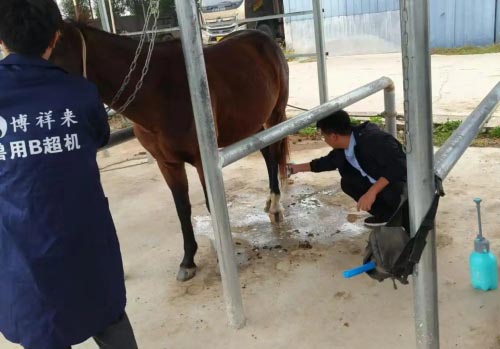
Portability, allowing veterinarians to perform field diagnostics
Monitoring capability, ideal for following healing progress over time
According to a 2022 review in Equine Veterinary Education, more than 70% of equine practitioners in the U.K. use ultrasound as a primary diagnostic tool for distal limb lameness.
Diagnostic Process: What Does Ultrasound Reveal?
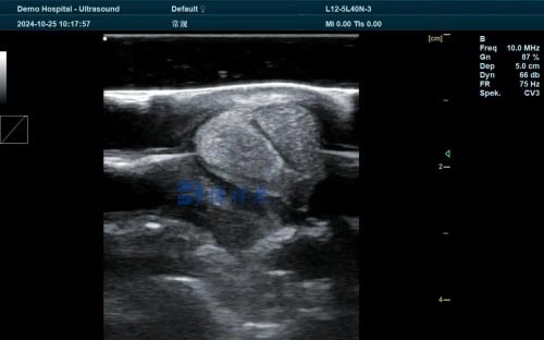
Using a linear high-frequency transducer—typically in the 7.5–15 MHz range—veterinarians can visualize equine tendon and ligament fibers in both longitudinal and transverse planes. A healthy tendon appears as a uniformly echogenic, fibrillar structure. Injuries present as hypoechoic (dark) or anechoic (black) lesions, representing fiber disruption, edema, or hematoma.
The following are commonly assessed:
Superficial Digital Flexor Tendon (SDFT): Commonly injured in racehorses and jumpers, often presenting with bowing and swelling.
Deep Digital Flexor Tendon (DDFT): Injuries can be harder to detect but may show up in the pastern or fetlock region.
Suspensory Ligament: Especially prone to proximal desmitis, particularly in dressage horses and eventers.
Annular Ligament & Check Ligaments: Assessed for impingement and associated tenosynovitis.
Veterinarians often score the injury based on lesion size, echogenicity, and fiber alignment. This grading system assists in prognostication and in tailoring rehabilitation protocols.
Ultrasound vs. MRI and Other Imaging
While Magnetic Resonance Imaging (MRI) offers a three-dimensional view of soft tissue and bone within the hoof capsule and deeper structures, its cost, need for anesthesia, and limited availability in field practice make it impractical for many equine veterinarians. Ultrasound, on the other hand, can be done stall-side, repeated frequently, and interpreted in real-time.
In comparative studies, ultrasound has been shown to detect over 85% of soft tissue injuries when compared to MRI in distal limb cases. For superficial injuries—particularly those in the metacarpal and metatarsal regions—ultrasound often yields diagnostic accuracy equal to or better than MRI in practical settings, especially when performed by skilled operators.
International Practices and Perspectives
Across Europe, North America, and Australasia, sport horse veterinarians use ultrasound as a cornerstone of lameness diagnosis. The Royal Veterinary College in London and the University of California, Davis have published protocols that emphasize standardization in image acquisition and interpretation.
One study published in Journal of Equine Veterinary Science (2021) highlighted that veterinarians in the Netherlands prefer using ultrasound not just for diagnosis, but for routine pre-purchase exams, particularly in older horses with high athletic demands. Similarly, Australian eventing veterinarians rely on portable ultrasound systems to conduct pre- and post-race assessments, identifying microtears that could evolve into more serious conditions.
Role in Rehabilitation and Prognosis
Ultrasound is not just a diagnostic tool—it’s a rehabilitation monitor. By documenting the healing process of tendons and ligaments over weeks and months, veterinarians can adjust rest and training regimens accordingly. This dynamic monitoring helps prevent re-injury, a common and frustrating outcome in sport horses.
In a 2023 study from the University of Guelph, ultrasound monitoring of the superficial digital flexor tendon during rehabilitation allowed clinicians to predict safe return-to-work timelines with 92% success. Horses monitored with routine ultrasound re-injury less frequently compared to those who returned to sport without structured follow-up imaging.
Limitations and Operator Dependency
Despite its advantages, ultrasound is highly operator-dependent. Poor image acquisition, incorrect probe placement, or misinterpretation can lead to misdiagnosis. Additionally, deep or intra-articular structures may be less visible compared to MRI or CT.
To address this, more veterinary schools and continuing education programs are offering advanced training in equine musculoskeletal ultrasonography, aiming to reduce variability and improve outcomes. Equipment quality also matters: newer portable machines like the BXL-V50 have improved resolution, depth penetration, and probe flexibility, making field diagnostics more accurate than ever before.
Future Directions: AI and Image Enhancement
Emerging technologies such as artificial intelligence (AI)–assisted interpretation, elastography, and 3D ultrasound reconstruction are beginning to enter the equine imaging field. These innovations could soon offer even greater diagnostic precision, reduce operator variability, and allow remote consultations using cloud-based platforms.
Some European equine hospitals are already trialing machine learning algorithms to automatically grade tendon lesions, improving diagnostic consistency and allowing quicker decision-making during sports seasons.

Conclusion
Ultrasound has proven itself as a powerful diagnostic tool in the detection and management of soft tissue injuries in sport horse limbs. It combines real-time, repeatable imaging with non-invasive safety, making it a preferred choice for equine veterinarians worldwide. While it may not replace MRI for complex or intra-articular injuries, its role as a front-line diagnostic and rehabilitation tool is unmatched in field practice.
With advancements in probe technology, imaging software, and AI-driven interpretation, the performance of ultrasound in equine sports medicine is only set to improve. For any veterinarian working with high-performance horses, mastering the use of ultrasound is not just valuable—it is essential.
References:
Whitton, R.C., et al. (2021). Ultrasound of the Equine Distal Limb: Diagnostic Protocols and Applications. Equine Veterinary Education.
Dyson, S.J., & Murray, R. (2022). Equine Lameness: Diagnosis and Management. Journal of Equine Veterinary Science.
van Weeren, P.R., & Crevier-Denoix, N. (2020). The Use of Ultrasonography in Equine Tendon Rehabilitation. Veterinary Radiology & Ultrasound.
Guelph Equine Performance Centre. (2023). Ultrasound Monitoring in Rehabilitation Protocols for Sport Horses.
Royal Veterinary College. (2022). Ultrasound Guidelines for the Equine Athlete.

