In the evolving world of animal agriculture, reproductive efficiency has become one of the most critical determinants of profitability. Fertility management, early pregnancy detection, and the diagnosis of reproductive disorders are central to ensuring that farm animals, particularly in the beef, dairy, sheep, and swine sectors, remain productive. One tool has proven itself indispensable in this arena: ultrasound technology.
While farmers continue to refine breeding strategies and enhance genetic selection, it is often the farm veterinarian—armed with real-time ultrasonography—who brings insight, precision, and timely intervention to the reproductive cycle. This article explores how veterinarians, especially in Western countries, use ultrasound to manage reproductive health on farms and why this non-invasive technology is increasingly considered the gold standard in livestock reproduction monitoring.
")
The Expanding Role of Farm Vets in Reproductive Management
Veterinarians have traditionally played a reactive role in reproduction—called in after failed inseminations or suspected pregnancy loss. However, with the widespread use of portable ultrasound, their involvement has shifted toward proactive herd fertility management. In countries like the United States, Canada, Australia, and across Europe, veterinarians are now routinely integrated into reproductive planning, using ultrasound to:
Detect pregnancies at the earliest possible stage
Monitor embryonic and fetal health
Evaluate ovarian and uterine status pre- and post-breeding
Identify reproductive tract pathologies
Aid in synchronization programs
This integration has significantly improved reproductive performance metrics such as calving interval, conception rate, and embryo survival—especially in dairy and beef cattle operations.
Ultrasound: A Vital Tool in Veterinary Reproductive Services
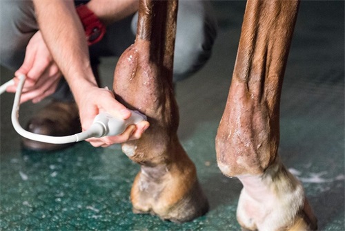
Ultrasound, particularly B-mode imaging, allows veterinarians to visualize reproductive structures in real-time. This includes the uterus, ovaries, and developing embryos or fetuses. In modern practice, ultrasounds are often performed transrectally in large animals such as cows and horses, and transabdominally in smaller species like sheep, goats, and pigs.
Some key reproductive evaluations that veterinarians perform using ultrasound include:
Pregnancy Diagnosis: In cattle, pregnancy can be diagnosed as early as 25 to 30 days post-insemination. In sheep and goats, it is typically performed between 35 and 45 days post-breeding. Early detection allows for rebreeding opportunities and herd fertility optimization.
Ovarian Activity Assessment: By evaluating the size and development of ovarian follicles and corpora lutea, vets can determine whether the female is in estrus, has ovulated, or is in a state of anestrus (no reproductive activity). This is crucial for timed artificial insemination (TAI) programs.
Embryonic and Fetal Viability Monitoring: Detecting a heartbeat, fetal movements, or placental abnormalities helps in assessing whether a pregnancy is progressing normally. Vets can also monitor twin pregnancies or assess fetal age, which aids in precise calving predictions.
Uterine Health: Postpartum ultrasound evaluation helps identify retained placenta, metritis, or endometritis, which if untreated, can impair future fertility.
The Shift Toward Preventative Reproductive Health
In North American and European herd health programs, ultrasound has become the core of preventative reproductive strategies. For instance, routine reproductive herd checks are now common in progressive dairy farms. These checks—performed weekly or biweekly by veterinarians—involve scanning a group of animals to track their reproductive cycle, confirm pregnancies, and diagnose any fertility disorders.
With these regular scans, farm vets can identify “problem cows” (e.g., those with silent heats, ovarian cysts, or uterine infections) before they cause extended open days (non-pregnant days), which are costly for producers. These interventions lead to:
Shorter calving intervals
Improved heat detection accuracy
Reduced reliance on hormonal treatments
Better culling decisions based on reproductive performance
Benefits of Ultrasound Over Traditional Methods
Compared to traditional techniques like rectal palpation, ultrasound offers numerous benefits:
Higher Accuracy and Early Detection: Ultrasound can confirm pregnancy earlier than palpation, with fewer errors, especially in young or small-framed animals.
Visualization of Structures: Vets can observe follicular waves, corpus luteum activity, and fetal development with much greater clarity.
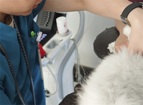
Non-invasive and Low Stress: With skilled handling, ultrasound causes minimal discomfort to the animal, making it ideal for repeated use.
Immediate Data for Decision-Making: Results are available in real time, allowing for instant reproductive planning adjustments.
Integration with Farm Technology and Recordkeeping
In technologically advanced livestock operations, veterinary ultrasound data is often integrated into herd management software. This ensures that all reproductive records—such as heat cycles, insemination timing, pregnancy status, and fetal age—are automatically stored and analyzed.
These integrations help farmers make informed decisions about:
Optimal insemination timing
When to dry off dairy cows
When to administer reproductive hormones
Replacement heifer selection based on reproductive traits
In particular, portable ultrasound machines such as the BXL-V50, designed for use in challenging farm environments, offer high-resolution imaging and long battery life. These features enable vets to perform multiple exams in the field without access to power, improving workflow and convenience.
Species-Specific Applications
1. Dairy and Beef Cattle:
In cows, reproductive ultrasound is used to confirm pregnancies, manage embryo transfer programs, and detect ovarian and uterine abnormalities. Some operations also use ultrasound for fetal sexing (between 55–80 days of gestation), which helps in planning for replacement heifers or bull calves.
2. Sheep and Goats:
Due to their seasonal breeding and high likelihood of twinning, ultrasound is indispensable in small ruminants. Farmers use the information for nutritional grouping—feeding twin-bearing ewes more than singles, which improves lamb birth weights and survival.
3. Swine:
In pigs, ultrasound is used to confirm pregnancy 25–30 days after mating. Monitoring uterine condition post-farrowing is also crucial in identifying complications like retained fetuses or postpartum infections.
4. Equine:
In mares, early pregnancy scanning can identify twins (which is dangerous in horses), allowing veterinarians to perform a "pinch" procedure to reduce one embryo. Ovulation timing, follicular development, and uterine health are also evaluated via ultrasound in breeding mares.
Training and Skill Development for Farm Vets
In Western countries, ultrasound has become part of standard veterinary curriculum and continuing education programs. Organizations like the American Association of Bovine Practitioners (AABP) and the British Cattle Veterinary Association (BCVA) offer training courses and certifications in reproductive ultrasonography.
Farmers increasingly rely on vets with ultrasound expertise, and in many cases, contract these services regularly during key breeding seasons. As such, the skill is both a clinical tool and a competitive advantage for veterinary professionals.
Challenges and Limitations
Despite its many advantages, ultrasound does come with limitations:
Operator Dependency: The accuracy of the exam depends heavily on the vet’s skill and experience.
Equipment Cost: High-quality ultrasound machines are expensive, though portable, farm-optimized models are becoming more affordable.
Access in Remote Areas: In some rural regions, limited veterinary access still hampers widespread use.
However, with advancements in wireless, battery-operated ultrasound units and telemedicine, these limitations are gradually diminishing.
Conclusion
For today’s farm vet, ultrasound is more than just a diagnostic tool—it’s a strategic instrument for reproductive management. Whether it’s identifying non-pregnant animals for rebreeding, confirming twin pregnancies in ewes, or monitoring uterine health post-calving, ultrasound allows for early, informed, and targeted interventions.
As global livestock industries push for higher reproductive efficiency and sustainable animal production, the integration of ultrasonography into routine veterinary practice continues to grow. From dairy barns in Wisconsin to sheep farms in New Zealand, ultrasound technology is helping veterinarians and farmers work smarter, ensuring healthy animals and profitable outcomes.
References
Whitaker, D. A., & Smith, E. (2021). Veterinary Ultrasonography in Food-Producing Animals. Journal of Veterinary Imaging.
Beef Cattle Institute. (2023). “Use of Ultrasound for Growth Evaluation in Cattle.” Retrieved from https://www.beefcattleinstitute.org/ultrasound-growth
American Association of Bovine Practitioners (AABP). (2024). “Ultrasound Reproduction Training for Practicing Veterinarians.” Retrieved from https://aabp.org
British Cattle Veterinary Association (BCVA). (2024). “ReproScan and Advanced Imaging Courses.” Retrieved from https://www.bcva.org.uk/training
link: https://www.bxlimage.com/nw/1260.html
tags: