Understanding the Role of Ultrasound in Exotic and Aquatic Animal Care
In modern veterinary medicine, the use of ultrasound imaging is expanding far beyond traditional farm animals and small domestic pets. One of the most fascinating and specialized applications of this technology lies in zoos and marine mammal facilities. Here, ultrasound plays a critical role in managing the health, reproduction, and overall welfare of a wide range of exotic species, from elephants and giraffes to dolphins and sea lions. For many veterinary professionals working with these animals, ultrasonography has become an indispensable, non-invasive diagnostic tool—offering unique insights into the internal state of species that are otherwise difficult or dangerous to assess.
")
This article explores how ultrasound machines are used in zoos and marine mammal environments, highlighting the challenges, innovations, and global veterinary perspectives that are shaping this specialized field.
Ultrasound in Zoo Animal Health Monitoring
Zoo animals represent an incredibly diverse group of species, each with unique anatomy and medical needs. Many zoo animals are also wild or semi-wild in behavior, making conventional physical exams risky or impractical. As a result, non-invasive tools like ultrasound are especially valuable.
Why Ultrasound Is Ideal for Zoo Animals
Non-invasive and safe: Unlike blood draws or surgical procedures, ultrasound causes no harm or stress when performed properly. For animals like gorillas, big cats, or rhinos, this is a major advantage.
Real-time internal imaging: It allows vets to visualize organs such as the liver, kidneys, heart, uterus, and lungs in real-time—crucial for diagnosing disease or monitoring pregnancy.
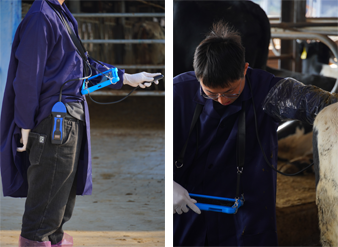
Minimally stressful with training: Many zoos now use positive reinforcement training to condition animals to accept ultrasound exams voluntarily. Elephants, for example, can be trained to present their belly or side for a scan without sedation.
Reproductive Monitoring and Pregnancy Detection
In many conservation programs, successful breeding is a key goal, particularly for endangered species. Ultrasound is widely used to:
Confirm pregnancy and determine fetal viability
Monitor uterine health in females
Track testicular condition and sperm production in males
Time ovulation for artificial insemination procedures
For example, ultrasound was used in the first successful artificial insemination of a female giant panda at the Smithsonian National Zoo, allowing the veterinary team to monitor fetal development without disturbing the mother.
Ultrasound Use in Marine Mammal Medicine
Marine mammals—dolphins, whales, seals, and sea lions—pose a unique set of challenges for diagnostic imaging. Their aquatic lifestyle, thick blubber layers, and large body sizes make many traditional imaging methods difficult. Fortunately, ultrasound has proven effective in many situations.
Unique Challenges of Marine Mammal Ultrasound
Water transmission: Ultrasound waves transmit differently in water, but with the right technique, scanning can be done directly through wet skin or submerged parts.
Thick skin and blubber: The probe needs to penetrate deeper tissues, requiring machines with high power and low-frequency probes for sufficient depth.
Limited restraint options: Most marine mammals are trained using operant conditioning to voluntarily participate in their own health checks, including ultrasound exams.
Common Applications in Marine Mammals
Pregnancy monitoring: Ultrasound is essential for confirming and monitoring pregnancy in bottlenose dolphins, beluga whales, and manatees.
Organ health assessment: Liver, kidney, and spleen diseases can be evaluated using abdominal scans.
Musculoskeletal imaging: Injuries in flippers or tails can be visualized using ultrasound, aiding in rehabilitation planning.
Guided procedures: In some facilities, ultrasound is used to assist in biopsies or guided fluid extraction.
Advances in Equipment and Portability
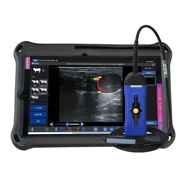
The development of portable and durable ultrasound machines, such as those used in rugged environments or field conditions, has significantly enhanced their usefulness in zoos and aquariums. Equipment must often be:
Battery-operated, with long runtime (especially in off-site or mobile settings)
Lightweight and waterproof, suitable for use near water or on boats
Flexible in probe design, offering both convex and linear transducers for varied anatomy
Able to store and export data, including still images and video loops for remote consultations
Veterinary professionals working with exotic and aquatic species frequently cite the need for user-friendly interfaces and real-time image quality as critical features. Many modern systems now integrate Doppler functionality to assess blood flow in uterine, cardiac, and vascular structures.
Case Studies and Global Best Practices
Case 1: Dolphin Pregnancy in Florida
At a marine mammal park in Florida, veterinarians used portable ultrasound equipment to monitor the pregnancy of a bottlenose dolphin over a 12-month period. By capturing weekly sonographic images, they successfully observed fetal growth stages and adjusted care protocols based on visualized fetal heart rate and activity. This close monitoring helped ensure a safe delivery and has since become standard practice.
Case 2: Elephant Reproductive Management in Europe
In several European zoos, elephants are trained for transrectal or transabdominal ultrasound scanning. Ultrasound allows zookeepers to detect ovarian follicles, time ovulation, and support artificial insemination programs—all of which are vital for sustaining captive populations. These efforts have contributed to increased breeding success among African and Asian elephants.
Case 3: Sea Lion Liver Disease in Japan
A large sea lion at a Japanese aquarium showed signs of lethargy and reduced appetite. Traditional blood work was inconclusive. Using a waterproof portable ultrasound scanner, vets were able to diagnose liver inflammation and administer early treatment—ultimately saving the animal. This illustrates the diagnostic precision that ultrasound can offer without requiring anesthesia or transport.
Collaboration with Technology Providers
Leading veterinary ultrasound manufacturers are increasingly collaborating with zoological institutions to customize machines for these unique needs. Features commonly requested include:
Preset settings for large and exotic species
Enhanced depth resolution for thick-bodied animals
Multilingual interfaces and telemedicine compatibility
In the field, devices like the BXL-V50—a waterproof, portable ultrasound scanner—are gaining popularity due to their durable design, intuitive controls, and long battery life, making them well-suited for use in zoos and marine parks.
Conclusion: A Growing Field with Global Impact
Ultrasound technology is playing a transformative role in the veterinary care of zoo and marine mammals. Its non-invasive nature, diagnostic accuracy, and compatibility with positive animal training methods make it uniquely valuable in these settings.
Around the world, veterinarians and researchers are harnessing ultrasound to improve breeding outcomes, diagnose complex diseases, and enhance the daily care of animals that were once considered too difficult to examine internally. As technology continues to evolve—and as our commitment to animal welfare grows—ultrasound will remain a cornerstone of responsible, science-based exotic animal care.
Whether it’s a pregnant dolphin swimming alongside the ultrasound probe or a gorilla voluntarily presenting her abdomen for a scan, these images not only reflect clinical success but also the growing trust and collaboration between humans and the animals in their care.
References
Smithsonian’s National Zoo. (2022). “Using Ultrasound in Panda Pregnancies.”
https://nationalzoo.si.edu/animals/news/ultrasound-gives-insight-panda-pregnancyNOAA Fisheries. (2023). “Marine Mammal Health Monitoring with Ultrasound.”
https://www.fisheries.noaa.gov/feature-story/ultrasound-monitoring-marine-mammal-healthEuropean Association of Zoos and Aquaria (EAZA). (2021). “Veterinary Imaging in Zoos: Best Practices.”
https://www.eaza.net/assets/Uploads/Files/Veterinary-Committee/Ultrasound-Zoos-Report.pdfVeterinary Radiology & Ultrasound Journal. (2023). “portable ultrasound in Aquatic Mammals.”
https://onlinelibrary.wiley.com/journal/17408261
link: https://www.bxlimage.com/nw/1259.html
tags: