In recent decades, veterinary ultrasound has emerged as a core tool in animal diagnostics, replacing guesswork with clarity across a wide range of species. Yet within this progress lies a striking contrast: the way we use ultrasound on large farm animals—like cows, horses, and pigs—is often vastly different from how it’s applied to small pets like dogs and cats. From the size of the equipment to the types of conditions we’re diagnosing, the divergence between large animal ultrasound and pet ultrasound reflects deeper differences in animal physiology, economic priorities, and even the environments where animals are treated.
")
In this article, we’ll explore the key distinctions between these two approaches—not just from a technical angle, but through the lens of practical use in real-life veterinary settings around the world. Whether you’re a livestock producer, a pet owner, or a vet in training, understanding these differences will help you better appreciate how this technology adapts to the needs of very different patients.
Understanding the Context: Large Animals vs. Companion Animals
At the heart of the matter is one basic truth: large animals and pets live vastly different lives and serve vastly different purposes. Large animals—cattle, sheep, goats, horses, pigs—are often raised for production: meat, milk, wool, breeding, or labor. Companion animals like dogs and cats are usually integrated into households, where their primary role is emotional and social.
This distinction heavily influences how and why ultrasound is used. On farms or breeding operations, the focus is often on reproductive efficiency, herd health, or quick assessment in outdoor environments. In small animal clinics, however, ultrasound is more commonly used for internal medicine, tumor screening, and emergency care.
Equipment Design and Portability
One of the most obvious differences lies in the design and use of the ultrasound equipment itself. Large animal practitioners need rugged, portable ultrasound systems that can be carried into barns, pens, or pastures. The machine has to work in dusty, wet, unpredictable environments and still deliver clear imaging.
Veterinarians working with large animals typically use handheld or wrist-mounted systems with protective casings. Some models, like the BXL-V50, are built for this type of work—offering waterproof designs, glove-friendly controls, and long battery life. This is crucial when scanning animals that may be nervous or difficult to restrain.
Pet ultrasound equipment, by contrast, is often larger and more stationary. These machines resemble those used in human medicine and are typically located inside veterinary clinics or hospitals. Here, the environment is controlled, the patient is likely sedated or calm, and image quality can be prioritized over mobility.
")
Patient Handling and Restraint
Another major difference between the two domains is how animals are handled during an ultrasound exam. Companion animals are often small enough to be lifted onto a padded exam table, where they can lie quietly on their sides. Vets or vet techs may gently restrain them while applying gel and moving the probe. Some dogs and cats are even trained to tolerate the procedure without sedation.
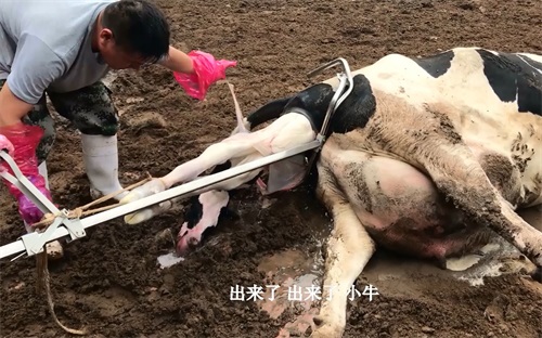
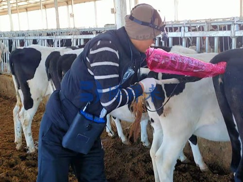
With large animals, things aren’t so simple. A dairy cow or mare is rarely going to lie down for an ultrasound. Exams are usually conducted with the animal standing, restrained in a chute or stocks, often with minimal sedation. This makes transrectal exams more common in large animals, especially for reproductive checks, since it's one of the most direct ways to access the uterus or ovaries.
In horses, rectal ultrasound is frequently used to diagnose colic or monitor pregnancy, while in cows, it’s used to assess uterine health, detect follicles, or confirm pregnancy. The operator often wears a full-length glove and manually guides the probe with one arm inserted into the rectum—a stark contrast to scanning a sleeping cat’s abdomen in a softly lit clinic.
Diagnostic Objectives and Economic Considerations
Another key distinction is why the ultrasound is being used in the first place. In large animal medicine, ultrasound is primarily an economic tool. It helps farmers make decisions that directly affect productivity: Is the cow pregnant? Is the sow ready to breed again? Is the racehorse suffering from a soft tissue injury?
Speed is essential. Large animal vets are often under pressure to move quickly, scanning many animals in one day. The images don’t have to be perfect—just good enough to make a clear decision. That’s why black-and-white B-mode imaging, not advanced Doppler or 3D, is often the go-to.
With pets, the stakes are different. Owners are often willing to pay more for detailed diagnostics, even if the pet isn’t economically “productive.” This opens the door for more complex ultrasound uses: diagnosing tumors, monitoring heart function with echocardiography, or performing guided biopsies. Here, the focus shifts from herd-level decisions to individual care.
Types of Ultrasound Examinations
Reproductive Scans
In large animals, reproduction is a primary application of ultrasound. Dairy farmers use it to track ovulation or confirm pregnancy. Breeders use it to check for twin pregnancies or fetal abnormalities. In swine, ultrasound helps monitor fetal number and gestational progress to optimize farrowing schedules.
In dogs and cats, reproductive ultrasound is often used for confirming pregnancy or estimating fetal viability—but it’s far less common. Many pet owners opt to wait and see, especially since smaller litters reduce economic urgency.
Abdominal Imaging
Abdominal ultrasound is common in pets and is used to assess the liver, spleen, kidneys, bladder, intestines, and other organs. It helps detect tumors, fluid buildup, or obstructions.
In large animals, abdominal ultrasound is more limited by size and depth. For example, it’s often used to check for displacement of the abomasum in cattle, or colic-related problems in horses. But it’s not typically used for full abdominal mapping like it is in cats or small dogs.
Musculoskeletal Scans
Horses are the primary large animal where musculoskeletal ultrasound is widely used. It's a key part of diagnosing tendon or ligament injuries, often in athletic or performance horses. This parallels small animal use in agility dogs or high-performance breeds.
")
Training and Technique
Large animal ultrasound requires a different set of skills. Operators must often work in the field, manage large animals safely, and interpret lower-resolution images in real-time. This places a premium on tactile feedback, anatomical familiarity, and speed.
In small animal practice, ultrasound is often performed by specialists or trained technicians. They have more time to review images, consult with radiologists, and use more advanced imaging modes.
Across both domains, training matters—but the technique adapts to the animal, the setting, and the purpose of the exam.
Case Example: On the Farm vs. In the Clinic
Consider this: A beef farmer in rural Nebraska uses a waterproof, portable ultrasound scanner like the BXL-V50 to check 60 cows for pregnancy in one morning. Each scan lasts less than two minutes, and decisions are made on the spot—breed again or cull.
Meanwhile, a veterinarian in an urban clinic sees a 12-year-old Labrador with lethargy and vomiting. She spends 20 minutes scanning the abdomen with a color Doppler machine to assess blood flow to a suspected liver mass. The image is sent to a veterinary radiologist for a second opinion. The owner gets a full report by the afternoon.
Both are ultrasound exams. But they couldn’t be more different.
Why the Difference Matters
Understanding these differences helps veterinarians, technicians, and animal owners make informed choices about equipment, technique, and expectations. It also shows how flexible and powerful ultrasound technology has become.
The future will likely bring more overlap. Portable color Doppler units may become common on farms. Clinics may adopt field-hardened models for mobile services. But the core distinction—between population-level management and individual animal care—will likely remain.
Ultrasound doesn’t just image organs—it reveals the practical priorities of the people who use it.
")
Conclusion
Ultrasound in veterinary medicine is not a one-size-fits-all solution. Whether scanning a high-producing dairy cow or a beloved housecat, the goals, tools, and techniques must fit the animal and the context. Large animal ultrasound values speed, durability, and decision-making on the move. Pet ultrasound leans toward precision, comfort, and long-term case management.
Both are vital. Both are evolving. And as technology advances, the gap between them may narrow—but the purpose behind each scan will always reflect the unique bond between humans and the animals they care for.
References
Whitaker, D. A., & Smith, E. (2021). Veterinary Ultrasonography in Food-Producing Animals. Journal of Veterinary Imaging.
Beef Cattle Institute. (2023). “Use of Ultrasound for Growth Evaluation in Cattle.”
https://www.beefcattleinstitute.org/ultrasound-growthAmerican College of Veterinary Radiology. (2022). “Ultrasound Guidelines for Small Animal Practice.”
https://www.acvr.org/small-animal-ultrasoundUniversity of Minnesota Extension. (2024). “Pregnancy Detection in Cattle Using Ultrasound.”
https://extension.umn.edu/reproductive-management/pregnancy-ultrasound