In the modern era of veterinary medicine, having the right tools is no longer a luxury—it’s a necessity. As more clinics and farms integrate technology into their workflows, ultrasound systems have emerged as one of the most essential diagnostic instruments for veterinarians. Whether you work with large livestock like cattle and pigs or small animals such as dogs and cats, the need for a high-performing ultrasound machine is universal. But with so many options on the market, how do you choose the best ultrasound for your veterinary practice?
")
This article will guide you through the key considerations when selecting a Veterinary ultrasound, the clinical and economic benefits it offers, and what international veterinary professionals look for when making this critical decision.
Understanding the Role of Ultrasound in Veterinary Practice
Veterinary ultrasound is a non-invasive imaging technology that allows real-time visualization of internal organs, muscles, blood flow, and reproductive status. It plays a vital role across species and clinical applications, including:
Pregnancy diagnosis
Reproductive management (e.g., follicular monitoring, ovulation timing)
Abdominal and cardiac imaging
Muscle and fat analysis in meat production animals
Detection of pathological changes (e.g., cysts, tumors, fluid accumulations)
Compared to X-rays, ultrasound does not use ionizing radiation, making it safer for repeated use—especially important in breeding and routine health monitoring.
Core Factors to Consider When Choosing an Ultrasound
1. Species and Application Scope
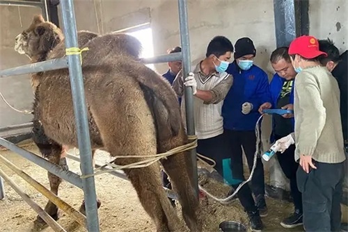
One of the first questions to ask is: What animals will you scan, and for what purpose? For large animals like cows, horses, and pigs, you’ll need an ultrasound with deeper penetration and possibly a linear or convex probe that accommodates transrectal scanning. In contrast, small animal practices may prioritize image resolution and compactness.
According to international studies (e.g., McLellan et al., 2022, Veterinary Radiology & Ultrasound), veterinarians working with multiple species often choose portable units that support interchangeable probes, allowing one machine to do the work of several.
2. Image Quality and Processing Technology
Image clarity is everything in diagnostics. A blurry scan could lead to misdiagnosis or the need for re-evaluation. In high-end veterinary practices, Doppler imaging and tissue harmonics have become the norm, enhancing vascular visualization and tissue contrast.
Advanced systems such as the BXL-V50 have earned praise for their combination of HD image resolution, long battery life (over 7 hours), and waterproof design (IP56), making them suitable for both clinical and field environments. These features are especially valued by veterinarians in Europe, Australia, and North America who often work in challenging weather or outdoor conditions.
")
3. Portability and Durability
Field veterinarians, especially those working on large farms or with wildlife, require a system that is lightweight, durable, and easy to disinfect. In countries like Canada and Argentina, where remote livestock inspection is common, wireless or handheld systems with rugged casings are preferred.
Battery life is another critical consideration. Many top-tier veterinarians opt for systems that can run for several hours without recharging, allowing for extended scanning sessions in areas with limited access to electricity.
Economic and Clinical Advantages of Ultrasound Use
1. Cost-Efficient Disease Detection
Early diagnosis leads to early intervention. For example, scanning a cow post-calving to detect retained placenta or uterine infection prevents complications that could delay conception or reduce milk yield. In swine production, early pregnancy detection using ultrasound avoids feed waste on non-pregnant sows.
A case study published by the Beef Cattle Institute (2023) highlighted that using ultrasound for monitoring fat and muscle deposition in beef cattle resulted in a 12–15% increase in overall profitability due to more accurate finishing decisions and reduced feed costs.
2. Improved Reproductive Outcomes
In reproductive management, timing is everything. In Europe and the U.S., many farms rely on real-time follicular tracking using B-mode ultrasound to determine the ideal insemination time. This not only increases conception rates but also minimizes hormone usage.
Moreover, the use of ultrasound in embryo transfer procedures has become a standard in advanced breeding programs. Accurate detection of corpus luteum and uterine tone provides a reliable assessment of recipient suitability.
3. Better Client Communication and Confidence
Many veterinarians in private small animal practices have noted that being able to show real-time ultrasound images to pet owners enhances trust and understanding. This visual evidence often leads to better compliance with treatment plans and follow-up recommendations.
Foreign Perspectives: What Global Vets Are Saying
Veterinarians across the world have shared consistent preferences when it comes to ultrasound selection. Here’s a brief overview:
United States: Veterinarians emphasize portability and software integration, as telemedicine and remote consultations become more common.
Germany and the Netherlands: High-quality image resolution and ergonomic design are priorities, particularly in specialized clinics.
Brazil and Argentina: Ruggedness and battery longevity are top considerations, as fieldwork dominates veterinary services.
Australia: Multi-species support is vital due to the common practice of mixed-animal medicine.
These regional preferences underline a global truth: a good veterinary ultrasound must adapt to various environmental and clinical demands.
")
Key Features That Define a Good Veterinary Ultrasound
Let’s summarize what veterinarians across species and borders value most in their ultrasound systems:
| Feature | Why It Matters |
|---|---|
| High-resolution display | Ensures accurate visualization of tissues and organs |
| Probe variety | Allows usage for transabdominal, transrectal, or cardiac exams |
| Long battery life | Critical for field use and power-limited areas |
| Water- and dustproof housing | Protects device in harsh barn or field conditions |
| Doppler capability | Enables blood flow analysis for advanced diagnostics |
| Lightweight and portable design | Enhances mobility and user comfort during long shifts |
| User-friendly software interface | Reduces learning curve and increases efficiency |
The Importance of Manufacturer Support and Training
When choosing a machine, you're not just buying a product—you're entering into a relationship with a brand. Reliable after-sales support, regular software updates, and technical training are invaluable. Western veterinary schools and continuing education platforms increasingly emphasize the importance of ultrasound proficiency, with many offering certification programs.
Veterinarians new to ultrasonography often benefit from choosing systems that offer built-in tutorials, intuitive menus, and compatibility with third-party teaching modules.
Real-World Use Case: A Mixed-Animal Vet in Australia
Dr. Helen Morris, a mixed-animal veterinarian in New South Wales, shares her experience:
“I use ultrasound every day—for cattle pregnancy checks, diagnosing pyometra in dogs, and even monitoring tendon injuries in horses. What made the biggest difference was getting a system that could keep up with me: rugged enough for the paddock, but precise enough for the clinic. My current unit is light, waterproof, and gives me a full workday without needing a charge.”
Her story mirrors that of many rural veterinarians around the globe who rely on versatile, dependable systems.
Conclusion: Invest in the Right Tool for the Right Future
Choosing the best ultrasound system for your veterinary practice is about more than just features—it’s about supporting better animal care, boosting economic efficiency, and positioning your clinic for future growth.
Ultrasound technology continues to evolve rapidly. Devices like the BXL-V50, combining practicality and precision, are setting new standards for veterinary imaging worldwide. By aligning your selection with your clinical needs, working environment, and species focus, you can ensure you make a sound investment that enhances diagnostics, treatment, and client satisfaction.
As the veterinary field becomes increasingly data-driven and technology-dependent, ultrasonography is no longer optional—it’s essential.
")
Reference Sources:
Beef Cattle Institute. (2023). “Use of Ultrasound for Growth Evaluation in Cattle.” https://www.beefcattleinstitute.org/ultrasound-growth
McLellan, L., Johnson, P., & Freeman, R. (2022). Veterinary Radiology & Ultrasound, 63(3), 315–326.
Whitaker, D. A., & Smith, E. (2021). Veterinary Ultrasonography in Food-Producing Animals. Journal of Veterinary Imaging.
link: https://www.bxlimage.com/nw/1256.html
tags: