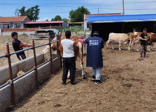
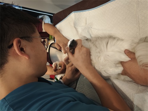
As a cattle producer, predicting when a pregnant cow will calve is essential for preparing appropriate care, ensuring safe delivery, and optimizing herd management. Traditionally, farmers have estimated calving dates based on breeding records, behavioral cues, or manual palpation, but veterinary ultrasonography—particularly B-mode ultrasound—has brought a new level of precision and confidence to this task. In this article, I’ll explore how veterinary ultrasonography is used to estimate the calving dates of cows, drawing from both practical experience and international research.

Understanding the Gestation Period in Cattle
The gestation period in cattle—the time from successful conception to the birth of the calf—typically lasts between 275 and 285 days, with an average of 280 days. However, this period can vary depending on several factors:
Breed: Early-maturing breeds tend to have slightly shorter gestation periods. Dairy breeds like Holstein often calve earlier than beef breeds like Angus or Charolais.
Sex of the Calf: Studies show that cows carrying heifer (female) calves usually have a gestation that is about 1 day shorter than those carrying bull (male) calves.
Age of the Cow: First-calf heifers often deliver slightly earlier than mature cows.
Season: Cows that are due in winter or spring may have longer pregnancies—by about 2–3 days—compared to those calving in summer or autumn.
Number of Fetuses: Twin pregnancies tend to have a shorter gestation period—by 3–6 days—than singletons.
Nutrition and Management: Poor nutrition or stressful conditions can delay parturition, lengthening the gestation period.
These nuances highlight the importance of individualized monitoring. While standard calendars provide helpful guidelines, ultrasound imaging can greatly refine the expected calving window.
Calculating Calving Date from Breeding Date
Once pregnancy is confirmed through ultrasound, the most common approach to estimating the expected calving date is to count 280 days forward from the breeding date. A widely used shortcut is the following:
Subtract 3 months from the breeding month.
Add 6 days to the breeding day.
For example:
A cow bred on May 2, 1986, would have an expected calving date of February 8, 1987 (May – 3 = February; 2 + 6 = 8).
A cow bred on May 2, 1987, would calve around February 6, 1988. In this case, you account for leap years by subtracting 1 day from the final result if February has 29 days.
This calculation provides a general prediction, but it doesn't account for individual variation, which is where ultrasonography becomes a powerful tool.
Role of Veterinary Ultrasonography in Pregnancy Monitoring
Veterinary ultrasonography provides a non-invasive, real-time view of the reproductive tract. It can detect pregnancy as early as 28 days post-breeding and allows veterinarians to assess fetal development, fetal age, and even estimate the likely calving date with increasing accuracy.
Fetal Age Estimation by Ultrasound
Between 30 to 150 days of gestation, ultrasonographic measurements of fetal size—such as crown-rump length, biparietal diameter (head width), and limb length—can be compared to established growth charts to estimate fetal age. This data helps back-calculate the conception date if the exact breeding time is unknown and refines the expected calving window.
International research has confirmed the high accuracy of this method:
A 2019 study from the University of Guelph, Canada, showed that fetal biometric measurements via ultrasound had a ±3–5 day error margin in cows between 60 and 120 days of pregnancy.
In Australia and New Zealand, where pasture-based systems demand efficient breeding schedules, ultrasound dating is essential for synchronizing calving and pasture availability.
Predicting Signs of Impending Parturition
As the calving date nears, ultrasonography also assists in identifying physiological signs that indicate an imminent birth. These include:
Mammary Development
About two weeks before calving, the cow’s udder begins to enlarge significantly. Within 2–3 days of parturition, the udder becomes tense and may show signs of redness and swelling. In some cows, colostrum leakage (pre-milk secretion) may be seen. Ultrasonography can detect tissue swelling and internal changes that signal milk accumulation and inflammation risk.Vulvar and Vaginal Changes
One week before calving, the vulva begins to swell and loosen. The mucous membranes turn pink and moist, and the cervical plug dissolves into a clear mucus that exits the vulva 1–2 days before delivery. Ultrasound helps assess the relaxation of the birth canal and confirms that the cow is approaching labor.Pelvic Ligament Relaxation
In the 7–10 days before calving, the sacrosciatic ligaments on either side of the tail head soften. This causes a noticeable depression near the base of the tail. In multiparous cows, this change is especially prominent. With ultrasound, the softening and spacing of pelvic structures can be visualized.Behavioral Changes
A cow nearing calving may isolate herself, show restlessness, lie down and stand up frequently, lift her tail, or look back at her abdomen. These behaviors, when combined with ultrasonographic findings, help pinpoint when labor is likely to begin.
Practical Application on the Farm
On our farm, we use Veterinary ultrasound at multiple stages of gestation:
30–60 days post-breeding: Confirm pregnancy, estimate age, and identify twins.
90–150 days: Use biometric measurements to verify fetal age and refine the calving calendar.
2 weeks prepartum: Monitor udder development, pelvic relaxation, and fetal positioning.
This allows us to:
Arrange for calving assistance if needed.
Avoid losses from unattended births.
Schedule vaccinations and nutritional supplementation at optimal times.
International Perspective and Adoption
In many Western countries, particularly the U.S., Canada, and parts of Europe, veterinary ultrasonography is considered a standard tool in reproduction management. It has become integral to the work of large-animal veterinarians and breeding consultants.
In developing regions, adoption is increasing thanks to the availability of portable, battery-powered ultrasound units. Organizations such as Heifer International and the Bill & Melinda Gates Foundation have supported the use of ultrasound to improve calving outcomes in rural herds.
Conclusion
Veterinary ultrasonography has revolutionized the ability to accurately estimate calving dates in cows. While traditional breeding records and observational methods are still valuable, ultrasound brings data-driven precision to pregnancy monitoring. By measuring fetal size, tracking tissue changes, and detecting signs of impending parturition, farmers can reduce calving risks, improve calf survival, and optimize herd productivity.
For any cattle operation—large or small—this tool has proven to be an indispensable ally in reproductive management. As awareness and access continue to grow, veterinary ultrasonography will likely become even more central to cow–calf operations around the world.
link: https://www.bxlimage.com/nw/1211.html
tags:


