As veterinary science advances, reproductive efficiency and precise monitoring of pregnancy in animals—especially livestock and companion animals—have become pivotal in optimizing breeding programs, pregnancy outcomes, and offspring health. Among the most reliable and non-invasive tools for tracking early pregnancy is ultrasonography focused on corpus luteum (CL) development. The CL, a temporary ovarian structure, produces progesterone essential for pregnancy maintenance. By measuring CL size, morphology, and blood flow using B-mode and color‑Doppler ultrasound, veterinarians can detect early pregnancies, assess luteal function, and predict pregnancy viability—helping both farmers and clinicians worldwide make informed decisions.
")
This article explores how Veterinary ultrasound is applied to track CL development and pregnancy across various species, highlighting recent foreign studies, instrumental techniques, and practical implications for monitoring fertility and early gestation.
1. The Role of Corpus Luteum in Pregnancy Maintenance
Following ovulation, granulosa cells differentiate to form the corpus luteum, which secretes progesterone to prime the uterus for embryo implantation and to prevent estrus recurrence. A well-functioning CL is vital: insufficient progesterone can lead to embryonic loss or failed implantation. While blood and milk progesterone monitoring have been employed, direct visualization and functional assessment of CL via ultrasound deliver real-time insights into luteal health.
Studies in dairy and beef cattle demonstrate that combining Doppler ultrasound analysis of luteal blood flow with progesterone levels allows identification of non-pregnant animals by Day 20–24 post-insemination. Another study in buffaloes showed that while CL size differed between pregnant and non-pregnant animals by Day 10 post-insemination, luteal blood flow and progesterone concentration earlier—around Day 7–8—served as more reliable early pregnancy indicators.
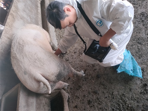
2. Veterinary Imaging Techniques
2.1 B‑Mode Ultrasonography
B-mode ultrasound records grayscale, cross-sectional images, allowing measurement of CL size and morphology. Veterinarians track diameters and volume growth—CL typically grows post-ovulation, reaches maximal size around Day 6–10, then regresses in non-pregnant animals. In dairy cows, CL volume began diverging between pregnant and non-pregnant groups as early as Day 9–10 .
2.2 Colour‑Doppler Ultrasonography
Doppler ultrasound visualizes luteal vasculature. Increased blood flow—quantified via blood flow area (BFA), time-average velocity (TAMV), or percentage area—reflects functional CL producing progesterone. Studies in buffaloes during Days 5–10 post-artificial insemination highlighted that pregnant animals showed significantly higher BFA/TAMV and progesterone levels than non-pregnant ones. In dairy cows too, CL blood flow at Day 20 perfectly predicted non-pregnancy, achieving 100% sensitivity in early diagnosis.
2.3 Combined Imaging and Hormone Assays
Multimodal approaches—combining ultrasonography with serum or milk progesterone—enhance diagnostic accuracy. Studies show that progesterone rises by Day 7, while blood flow signals detectable CL function by Day 8 . The combination enables differential diagnostics between physiological luteal phases and early luteolysis.
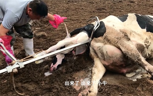
3. Applications Across Species
3.1 Cattle (Beef and Dairy)
In bovine reproduction, early pregnancy detection shortens calving intervals and improves reproductive planning. Ultrasound detection of CL volume and blood flow around Days 9–14 post-insemination predicts pregnancy success. Functional CL identification supports decisions for resynchronization or early embryo transfer. A review of cattle CL confirms the effectiveness of Doppler ultrasonography in assessing luteal activity in timed-insemination protocols.
3.2 Buffalo
Similar trends appear in buffaloes—even seasonal breeders—where CL development and vascularity patterns during Days 5–10 post-insemination significantly correlate with pregnancy outcomes.
3.3 Canines (Dogs)
In dogs, the CL is crucial to pregnancy maintenance: progesterone from CL remains high until term. A recent observational study on 26 bitches reported CL diameter increases post-ovulation correlated with progesterone, peaking mid-estrus, and plateauing after Day 35. Furthermore, CL visualization helps distinguish physiological cavitary CL from pathological cysts—improving ovulation timing and luteal health monitoring .
")
4. Why Ultrasound Is Gaining Ground in Veterinary Reproduction
Non-invasive and stress-free
Ultrasound enables frequent longitudinal monitoring without harming animals—critical for livestock welfare and intensive reproductive management.Quantitative and objective data
It provides precise measures of CL size, vascularity, and morphological changes, surpassing subjective palpation.Earlier diagnosis
Doppler ultrasound can detect functional CL activity earlier than progesterone assays—by Days 8–10—allowing faster reproductive synchronization.Species versatility
Applicable across cattle, buffalo, small ruminants, and companion animals like dogs.Economic and reproductive efficiency
Early identification of non-pregnant animals enables efficient management: rebreeding, early embryo transfer, or culling, reducing costs and improving herd productivity.
5. Workflow of CL‑Based Pregnancy Monitoring
Synchronization or breeding event (e.g. timed AI).
Schedule ultrasound days post-ovulation, typically Days 5–10.
Perform B-mode scan to record CL size, morphology.
Conduct colour‑Doppler scan to assess vascular parameters, like BFA or TAMV.
Optional blood/milk progesterone assay for hormonal context.
Interpret combined data:
Pregnant animals show enlarging CL, sustained vascularity, elevated progesterone.
Non-pregnant show early luteal regression: reduced blood flow without progesterone support.
Make decisions: re-synchronization, embryo transfer, or retention.
6. Challenges and Considerations
Equipment and training
Requires high-resolution ultrasound and trained operators. Inter-operator variability can impact quantitative readings.Species-specific timing
Cattle and buffalo show luteal patterns in Days 5–10; dogs show CL plateau till Day 35.Luteal anomalies
Presence of cavitary vs homogeneous CL in cattle raises questions on function; most evidence supports similar progesterone output.Environmental factors
Breed, nutrition, and management systems can influence luteal dynamics.
7. Future Perspectives and Research
Automated image analysis
Software (e.g. ImageJ) quantifies blood flow areas to improve diagnostic precision.Biomarker integration
Combining imaging with gene expression (e.g. ISG15, MX2) may refine early pregnancy diagnostics.Cross-species comparisons
Expanding comparative studies across livestock and companion species to standardize protocols.Portable devices
Development of cost-effective, handheld Doppler units could democratize reproductive monitoring on small farms.
Conclusion
Measuring corpus luteum development via veterinary ultrasound—especially using both B‑mode and color‑Doppler modalities—provides a powerful, non-invasive method for early pregnancy detection and luteal function assessment across species. With accurate insights by Days 8–10 post-breeding, veterinarians and farmers can make data-driven decisions to improve reproductive efficiency, reduce economic losses, and better manage animal welfare. As imaging technology and biomarker analysis evolve, ultrasound-based CL monitoring will increasingly become a standard in reproductive management for both livestock and companion animals.
")
References
Bó GA, et al. "Current status of corpus luteum assessment by Doppler ultrasonography in dairy and beef cattle." Animal 2023. DOI:10.1016/j.animal.2023.100752
Andrade JPN, et al. "Early pregnancy diagnosis in buffaloes using CL size, blood flow and progesterone." Frontiers in Veterinary Science 2020. DOI:10.3389/fvets.2020.00299
Paganotto A, et al. "Ultrasound monitoring of CL morphological evolution and serum progesterone in dogs." Veterinary and Animal Science 2025. DOI:10.1016/j.vas.2025.100444
De Silva M, et al. "Early pregnancy diagnosis in cows via CL blood flow analysis." BMC Veterinary Research 2024. DOI:10.1186/s12917-024-04438-5
Buffalo CL development and Doppler study. MDPI Animals 2019. DOI available
Mebarki Bovini Review: "The utilization of B‑mode and Doppler ultrasound in ruminants." Large Animal Review 2024