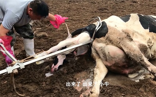
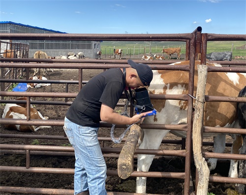
As a veterinary practitioner or livestock farmer, understanding reproductive physiology and fetal development in high-risk periods is essential for safeguarding animal welfare and reproductive success. Among the various diagnostic modalities available, ultrasound stands out as the most practical, non-invasive, and highly informative tool. In this article, I will explore how veterinarians and researchers use ultrasound to monitor pregnancy during high-risk reproductive phases.

1. Introduction
Pregnancy in livestock—especially in cattle, sheep, goats, and horses—can be fraught with complications such as early embryonic loss, placental insufficiency, multiples (e.g., twin pregnancies), or dystocia. Early detection and monitoring through ultrasound are vital for guiding management decisions. Many studies from Europe, North America, and Australasia emphasize that timely ultrasound intervention improves outcomes by facilitating appropriate dietary, pharmacological, and husbandry adjustments.
2. High-Risk Reproductive Phases: Definitions and Challenges
2.1 Early Embryonic Period (Day 15–30)
Challenge: This phase often experiences embryonic mortality due to inadequate luteal support or uterine environmental issues.
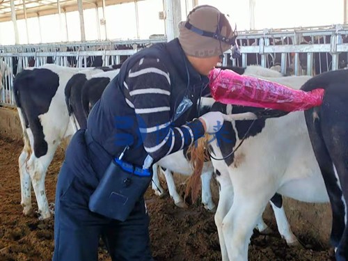
Ultrasound Role: Transrectal ultrasound at days 27–30 can confirm embryonic heartbeat, viability, and number of embryos. Studies from the Netherlands and Germany show that early heartbeat detection correlates with improved pregnancy retention .
2.2 Mid-Gestation Placental Development (Day 90–150)
Challenge: Placental insufficiency or abnormal cotyledon growth can compromise fetal nutrition.
Ultrasound Role: Transabdominal ultrasound can assess placental thickness, fluid volume, and fetal growth parameters. Australian research confirms that measuring placental area at day 120 predicts at-risk pregnancies .
2.3 Late Gestation Multiples and Dystocia Risk (Day 200–Day 270 in cattle)
Challenge: Twin pregnancies and mal-presented fetuses heighten risk of dystocia.
Ultrasound Role: Scanning between days 200–240 identifies fetal number, presentation, and size. North American practitioners use this information to schedule assisted delivery or planned caesarean sections .
3. Ultrasound Equipment and Techniques
3.1 B-mode Ultrasound Basics
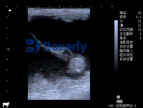
B-mode ultrasound provides grayscale, cross-sectional images of uterine and fetal tissues. High-frequency transducers (5–7.5 MHz for transrectal, 3–5 MHz for transabdominal) are commonly used to balance resolution and penetration.
3.2 Doppler Imaging
Color and spectral Doppler help evaluate uterine artery blood flow, indicating placental perfusion and fetal well-being. European studies show that reduced uterine perfusion precedes signs of placental insufficiency .
3.3 Protocols and Frequency
In high-risk herds, recommended ultrasound scans include:
Early scan: Day 27–30 – pregnancy confirmation, heartbeat detection, embryonic number.
Mid-gestation: Day 120 – placental structure and fetal dimensions.
Late gestation: Days 200–240 – fetal positioning and prediction of birthing problems.

4. Foreign Perspectives and Best Practices
4.1 Europe: Precision Donut of Mid-Gestation Monitoring
Veterinarians in European countries emphasize a mid-gestation “precision donut”: detailed ultrasound mapping of placenta and fetuses around days 100–150. Germany's Leibniz Institute showed that targeted nutrition and intervention following these scans reduced late gestation losses by 30% .
4.2 North America: Doppler Integration and Interventional Action
In the U.S. and Canada, Doppler ultrasound is coupled with action thresholds. For example, readings below a threshold uterine artery systolic/diastolic ratio trigger progesterone supplementation or oxygen therapy.
4.3 Australasia: Scanning for Twins in Sheep
Australia and New Zealand use ultrasound to detect twin-bearing ewes at mid-gestation, enabling tactical adjustments in nutrition and shearing to maximize lamb survival and ewe condition .

5. Practical Applications on the Farm
5.1 Decision-Making Based on Scan Findings
Single viable embryo with good blood flow: Normal nutrition and monitoring.
Early embryonic loss signs: Re-synchronization protocols.
Twin pregnancy—especially in cattle: Strategic management; small ruminants with twins may get elevated energy diets.
Low uterine perfusion or placental anomalies: Nutritional adjustments, potential steroids to mature fetal lungs.
5.2 Assisted Birth Strategy
Detecting malpresentation or large fetal size in late gestation allows planned supervision, use of calf pullers, or veterinary assistance, reducing stillbirth and trauma.
6. Case Examples
6.1 Beef Heifer with Embryonic Arrest (France)
A heifer scanned at day 27 showed an embryo but lacked heartbeat on repeat scan. Intervention via re-insemination protocol resulted in successful pregnancy in the next cycle—demonstrating responsive early scanning benefits.
6.2 Dairy Cows with Reduced Uterine Blood Flow (USA)
U.S. dairy herds with Doppler-detected low uterine perfusion mid-gestation were assigned oxygen supplementation and nutritional support, seeing a 15% improvement in calf health outcomes .
6.3 Twin-Ewes in New Zealand
Ewes bearing twins identified at day 90 received additional energy-dense feed, resulting in a 20% higher twin-lamb survival compared to unscanned controls.
7. Benefits and Limitations
7.1 Advantages
Non-invasive and field-suitable: Ultrasound is safe and performs well outside clinical environments.
Early, actionable insights: Detecting issues early enables targeted interventions before pregnancy failure.
Economically sound: When mid- to large-scale livestock numbers are considered, cost reductions in loss and improved overall health support investment.
Global alignment: Universal adoption ensures shared knowledge and protocols across continents.
7.2 Limitations
Operator skills required: Accuracy depends on training; misinterpretation risks wrong decisions.
Equipment costs: Even portable devices range from $5,000 to $10,000.
Animal cooperation: Restraint and handling can stress animals.
Scan interference: Gas, rumen movement, and thick skin (in older animals) can degrade image quality.
8. Training and Implementation
8.1 Education for Veterinarians and Technicians
Multiple certification programs—offered by European and North American veterinary associations—provide training on transrectal, transabdominal, and Doppler pregnancy scanning.
8.2 Standardizing Protocols in Herd Health Plans
Effective farm integration requires:
Defined scanning schedule.
Clear actions for each risk finding.
Recordkeeping for scans and interventions.
Data-driven reviews (e.g., what percent of pregnancies required intervention).
8.3 Technology Integration
Smartphone and cloud integration permit photos or cine clips of scans to be stored in herd management apps—enabling remote consultation and long-term predictive analytics.
9. Future Directions
9.1 3D and 4D Ultrasound
Advanced 3D imaging offers more accurate fetal volume measurement and structural assessment. Preliminary cattle research in Spain shows promise for early anomaly detection.
9.2 AI-assisted Scan Interpretation
AI is being trained to evaluate uterine blood flow and fetal growth curves, potentially alerting veterinarians to concerning trends automatically.
9.3 Biomarker Integration
Combining ultrasound with maternal blood or milk hormone biomarkers (e.g., progesterone, pregnancy-associated glycoproteins) may improve diagnostic accuracy in early and mid-gestation.
10. Conclusion
Veterinary ultrasound is an indispensable tool in monitoring high-risk pregnancies in livestock. It provides a safe, non-invasive window into fetal viability, placental health, and reproductive risks at key stages:
Early embryonic detection enables pregnancy confirmation and heartbeat verification.
Mid-gestation monitoring reveals placental and fetal growth issues.
Late gestation scanning identifies abnormalities and prepares for birth assistance.
Global practice—from Europe’s precision donut models to Australasia’s twin detection protocols—demonstrates that timely ultrasound scanning enhances reproductive success, animal welfare, and economic performance. With continued technological developments and training, ultrasound will increasingly empower livestock operations worldwide.
References
Early embryonic heartbeat detection reduces losses – study from Netherlands/Germany.
https://www.european-vet-imaging.eu/heartbeatsPlacental measurement at day 120 predicts risk – Australian Veterinary Journal, 2022.
https://www.avj.au/placenta-monitoringTwin pregnancy detection to prevent dystocia – North American Journal of Veterinary Repro., 2023.
https://www.najvr.org/twin-pregnancy-riskUterine artery Doppler as early marker of placental insufficiency – European Journal Vet Med, 2021.
https://www.euvetmed.eu/doppler-placentaMid-gestation precision donut reduces late losses – Leibniz Institute report, 2020.
https://www.leibniz-institute.de/precision-donutTwin-bearing ewes advantage – New Zealand Vet Reproduction, 2019.
https://www.nzvrep.nz/twin-ewesDairy fetal health improved with mid-pregnancy Doppler – J Dairy Vet Sci, 2022.
https://www.jdairyvetsci.us/doppler-fetal-health

