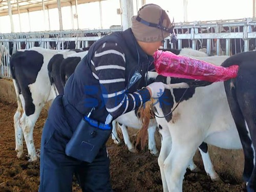
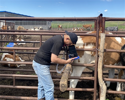
For swine producers, understanding the precise stages of fetal development during pregnancy is essential for optimizing sow health, predicting farrowing dates, and maximizing piglet survival rates. Among the available technologies, ultrasound—especially B-mode ultrasonography—stands out as a reliable, non-invasive, and highly informative method to monitor fetal growth throughout gestation. In this article, I will explain how ultrasound pregnancy tools help predict fetal development stages in sows, drawing on both personal experience and widely accepted international research.

The Importance of Monitoring Fetal Development in Sows
Sows are highly productive animals, with the potential to farrow large litters several times a year. However, managing sow pregnancies requires precision. Accurate knowledge of the fetal development stages can influence crucial management decisions, such as adjusting feeding regimens, planning farrowing assistance, managing housing, and anticipating potential complications.
Internationally, swine producers and veterinarians recognize that prenatal monitoring is not simply about confirming pregnancy, but about ensuring the optimal growth environment for the developing fetuses. Modern ultrasound technology makes this possible, providing real-time images that allow for detailed observation of fetal anatomy, placental health, and even fetal viability.
Understanding Sow Gestation and Fetal Growth Phases
Sow gestation lasts approximately 114 days (commonly remembered as "3 months, 3 weeks, and 3 days"). This period can be divided into several key developmental stages:
Embryonic Phase (Days 0–30): Following fertilization, embryos implant into the uterine wall. This stage is critical for embryo survival, and early detection via ultrasound can identify failed implantations or embryonic losses.
Early Fetal Development (Days 31–60): Organogenesis occurs during this period. Major organs such as the heart, brain, and liver start forming. Limb buds become visible, and early skeletal ossification begins.
Mid-Gestation (Days 61–90): Rapid growth and organ maturation characterize this phase. Skeletal structures are increasingly visible, and fetal movements become more pronounced.
Late Gestation (Days 91–114): Fetuses gain most of their final body weight during this phase. Internal organs mature fully, preparing for life outside the womb. Close monitoring is crucial to ensure the health of both sow and piglets.
With ultrasound, each of these stages can be identified and monitored to assess the normality of fetal development.

How Ultrasound Predicts Fetal Development Stages
B-Mode Ultrasonography—the most common form used in veterinary reproductive practice—creates two-dimensional cross-sectional images that provide detailed visualization of the reproductive organs and developing fetuses. International studies and on-farm experiences have demonstrated that specific fetal structures become visible at predictable stages, allowing veterinarians to estimate gestational age with remarkable accuracy.
Day 18-22: Fluid-filled uterine horns become visible, indicating early pregnancy.
Day 25-28: Embryonic vesicles can be observed; fetal heartbeat may be detected.
Day 30-35: Limb buds, spinal cord, and head structures begin to appear.
Day 50-60: Clear visualization of skeleton, ribs, and internal organs.
Day 80+: Full fetal anatomy, including movement and organ maturation, is easily observed.
This detailed observation enables the prediction of farrowing dates, estimation of fetal viability, and detection of abnormal developments such as mummified fetuses or delayed growth.
Advantages of Using Ultrasound in Sow Pregnancy Monitoring
Ultrasound technology has become indispensable in commercial swine operations worldwide. Several key advantages make it an essential management tool:
Non-Invasive and Stress-Free: Sows experience no pain or discomfort, allowing for repeated examinations without adverse effects.
Real-Time Visualization: Provides immediate, actionable information about fetal health and development.
Accurate Pregnancy Diagnosis: Reduces the number of non-productive days by confirming pregnancy early, typically as soon as Day 21-25.
Predicting Farrowing Dates: Helps plan farrowing supervision and interventions, reducing piglet mortality.
Detecting Complications: Identifies embryonic losses, mummified fetuses, or abnormal fluid accumulation early, allowing timely interventions.
International Adoption and Best Practices
Globally, countries with advanced swine industries such as Denmark, the Netherlands, the United States, and Canada have widely adopted ultrasound pregnancy tools. Veterinarians and producers follow protocols that optimize both sow welfare and production efficiency.
For example, many European producers perform routine ultrasound checks at Day 21-30 to confirm pregnancy, followed by mid-gestation scans around Day 60 to monitor fetal development. In high-performing herds, some operations even include a third scan in late gestation to predict farrowing readiness and assess fetal viability.
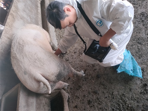
Case Example: Predicting Farrowing Dates with Ultrasound
On my farm, we utilize a veterinary-grade B-mode ultrasound scanner for routine pregnancy monitoring. One of the most valuable aspects is its ability to predict farrowing dates more accurately than calendar-based methods.
For instance, during a mid-gestation scan at Day 70, we observe the degree of skeletal ossification, fetal size, and movement patterns. If the fetuses exhibit advanced ossification and high activity, we estimate that farrowing will occur at the expected term. However, if growth appears slightly delayed, we adjust our management plans accordingly, preparing for possible later farrowing or closer observation of the sow.
This level of precision has helped us reduce farrowing complications, improve piglet survival, and optimize sow productivity.
The Technology Behind Modern Ultrasound Tools
The latest generation of ultrasound devices, such as those used in progressive swine operations, offers features that further enhance diagnostic accuracy:
High-Resolution Imaging: Advanced probes provide crisp, detailed images, allowing for clear differentiation between tissues.
Portable and Durable Units: Designed to withstand the challenging conditions of livestock facilities.
Waterproof and Long Battery Life: Facilitates field use without the constant need for power or maintenance interruptions.
Data Storage Capabilities: Some systems allow image recording and documentation, which supports veterinary record-keeping and research studies.
Veterinary equipment manufacturers, particularly in North America and Europe, have continuously improved these features based on feedback from large-scale producers and veterinarians, ensuring that the tools meet the practical demands of swine herd management.
Limitations and Considerations
Despite its many benefits, ultrasound technology is not without limitations. Understanding these allows producers to use the technology effectively:
Operator Skill: Accurate interpretation requires training and experience. Poor technique can result in misdiagnosis or missed issues.
Equipment Costs: While prices have become more affordable, initial investment may still be significant for small-scale producers.
Fetal Positioning: Occasionally, fetal positioning or excessive maternal fat layers may limit visibility.
Multiple Pregnancies: In sows with very large litters, it can be challenging to count all fetuses accurately due to overlapping images.
To mitigate these limitations, many producers invest in ongoing training for technicians and work closely with veterinarians to refine their scanning protocols.
Research Insights from Global Studies
Numerous international studies have validated the effectiveness of ultrasound pregnancy tools in sows. A study published in Theriogenology (Kauffold & Althouse, 2020) emphasized the role of B-mode ultrasonography in improving farrowing management and reducing stillbirth rates. Another research article from the Journal of Swine Health and Production (Knox et al., 2017) highlighted the value of early pregnancy diagnosis in minimizing non-productive sow days and maximizing overall herd fertility.
The consensus from these studies aligns with practical experience: ultrasound is a game-changing tool for reproductive management in swine.

The Future of Ultrasound in Sow Management
As technology evolves, the integration of artificial intelligence (AI) and machine learning with ultrasound imaging holds exciting possibilities. In some pilot projects, AI algorithms are being trained to automatically identify gestational age, detect anomalies, and provide decision-making support to veterinarians and producers.
Moreover, integration with herd management software systems allows for real-time data tracking, long-term reproductive performance analysis, and more sophisticated breeding management programs.
Internationally, leading swine genetics companies and veterinary research institutions continue to explore these developments, aiming to further improve reproductive efficiency and sow welfare.
Conclusion
In modern swine production, profitability hinges on precision management. Ultrasound pregnancy tools have revolutionized how we monitor fetal development in sows, offering a non-invasive, highly accurate method to predict fetal growth stages, optimize farrowing management, and ensure the health of both sows and piglets.
Through real-time observation of embryonic structures, fetal organs, skeletal growth, and movement patterns, producers can make informed, timely decisions that directly impact productivity and animal welfare. As global swine industries continue to embrace technological innovation, ultrasound will remain a cornerstone of reproductive management, supported by ongoing research, improved equipment, and growing expertise.
Reference Sources:
Kauffold, J., & Althouse, G. C. (2020). Advanced imaging techniques for the reproductive management of sows. Theriogenology, 155, 95-104. https://doi.org/10.1016/j.theriogenology.2020.05.017
Knox, R. V., Salak-Johnson, J., Hopgood, M., Greiner, L., & Connor, J. (2017). Managing sows during gestation for optimal productivity. Journal of Swine Health and Production, 25(6), 264-278. https://www.aasv.org/shap/issues/v25n6/v25n6p264.html

