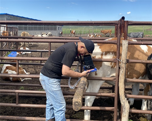
In the world of modern animal husbandry, timely and accurate pregnancy detection in livestock is a vital factor in improving reproductive efficiency, reducing economic losses, and optimizing herd management. Among the various diagnostic tools available, veterinary ultrasound—especially during the early gestation period—has emerged as one of the most trusted methods for confirming pregnancy. With advancements in imaging resolution, probe design, and software integration, the accuracy of ultrasound in early pregnancy detection has significantly improved, making it a standard practice in countries like the United States, Canada, Australia, and parts of Europe.

In this article, we will explore the accuracy of veterinary ultrasound pregnancy detection during early gestation, discuss the biological and technical factors that influence it, and examine how farmers and veterinarians worldwide use this non-invasive tool to support breeding decisions.
Understanding Early Gestation in Livestock:
The early gestation period refers to the first few weeks following successful fertilization. In cows, this generally includes days 25–45; in sheep and goats, it’s around days 20–40; in pigs, days 21–35. It is during this phase that embryo implantation, placental development, and early fetal growth occur. However, because the embryo is still very small and fragile, detecting it accurately requires high-sensitivity tools and skilled operators.
Foreign livestock producers emphasize early detection for several reasons:
Timely rebreeding: If a female is not pregnant, detecting this early allows the producer to reintroduce her into the breeding cycle.
Economic savings: Early detection prevents wasted feed, space, and labor on non-productive animals.
Health monitoring: Early scanning helps identify embryonic loss or uterine abnormalities.
How Ultrasound Works in Early Pregnancy Detection:
Veterinary ultrasound works by emitting high-frequency sound waves into the animal’s body using a probe. These waves reflect off tissues, and the returning echoes are converted into images on a screen. In early gestation, technicians look for anechoic (black) fluid-filled sacs indicating the presence of amniotic fluid and the embryo.
Two major methods are used:
Transrectal scanning: Common in cattle, it involves inserting a probe into the rectum to visualize the uterus. This method is highly accurate between days 28–45.
Transabdominal scanning: Common in small ruminants and pigs. The probe is applied to the abdomen, typically after day 25, depending on species and body size.
In countries like the U.S. and Australia, portable B-mode scanners with linear or convex probes are frequently used for early pregnancy checks in both confined and open-range conditions.
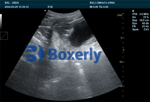
Accuracy of Ultrasound in Early Gestation
The accuracy of ultrasound pregnancy detection varies with species, gestation day, technician experience, and equipment quality. However, several studies and industry surveys provide valuable benchmarks:
Cattle: Around day 28–35, ultrasound accuracy is typically 95–98%. False negatives can occur before day 28 due to late implantation or small embryo size.
Sheep and Goats: Starting from day 25, accuracy reaches 90–95%, improving after day 30. Abdominal fat and rumen gas can interfere with imaging in overweight ewes or does.
Pigs: Transabdominal ultrasound has around 90–97% accuracy from day 23 onward. Earlier checks (before day 20) are more prone to error due to small fluid volume.
Importantly, foreign producers often highlight that repeat scans after 7–10 days increase overall accuracy and help detect early embryonic loss, which can otherwise go unnoticed.
Factors Affecting Accuracy:
Gestation Age
Detecting pregnancy too early (e.g., before day 25 in cows or pigs) may lead to false negatives due to insufficient fluid accumulation or undeveloped fetal structures.Operator Skill
In countries with formal training programs (like Canada or Denmark), technicians are trained to detect subtle early signs—like the echogenic ring of the embryo—reducing misdiagnosis.Equipment Quality
High-resolution ultrasound systems such as BXL-V50 or Mindray DP-30 are preferred. Devices with color Doppler help distinguish blood flow around the placenta, improving confirmation.Animal Condition
Factors like excessive body fat, full rumen (in ruminants), or uterine abnormalities may reduce image clarity. It’s common practice in some U.S. farms to restrict feed before scanning to improve visualization.
Benefits of Early Pregnancy Detection Using Ultrasound:
Economic Efficiency: Quickly removing non-pregnant animals improves space usage and reduces feed waste.
Reproductive Planning: Synchronizing rebreeding cycles reduces calving/kidding/lambing intervals.
Health Management: Abnormal fluid patterns or uterine inflammation can be identified early.
Animal Welfare: Ultrasound is non-invasive, causes no stress, and can be performed on-farm.
A study published by the Journal of Dairy Science (2020) emphasized that farms implementing early ultrasound checks had 12% higher pregnancy rates and saved an average of $38 per head annually due to improved reproduction efficiency.
Real-World Use Cases Around the Globe:
United States:
Dairy producers in Wisconsin and California routinely conduct transrectal scans around day 30 post-AI. This helps them rebreed open cows by day 45, maintaining compact calving seasons.
Australia:
In sheep stations across New South Wales, transabdominal scanners are used as early as day 25 post-breeding. Early detection allows producers to sort pregnant and non-pregnant ewes for targeted feeding.
Spain and Portugal:
Goat herds in Mediterranean climates use portable B-mode scanners for early fetal detection and number of fetuses, allowing better nutritional planning in late gestation.
China:
Swine farms using BXL-DB20 ultrasound machines confirm pregnancy in sows around day 25–30, optimizing batch farrowing systems.
Challenges and Limitations:
While ultrasound is widely effective, it does have some limitations:
Cost: Advanced devices can be expensive for small-scale farmers, though portable units are becoming more affordable.
False Positives: Fluid from uterine infections may resemble pregnancy sacs if not interpreted correctly.
Embryonic Loss: Pregnancy detected at day 28 may be lost by day 40, leading to incorrect assumptions without follow-up.
Veterinary best practice in North America recommends follow-up scans around day 45–55 to confirm fetal viability.
Conclusion:
Veterinary ultrasound has revolutionized early pregnancy detection in livestock, offering high accuracy, speed, and cost-effectiveness. When performed correctly and at the appropriate time, it allows producers to make informed decisions about breeding, feeding, and herd management. International experience shows that ultrasound not only boosts reproductive success but also improves the economic sustainability of livestock operations.
As ultrasound devices become more affordable, durable, and portable, we can expect their use to become even more widespread across diverse farming systems. With proper training and timing, early gestation scanning provides farmers with reliable insight into the hidden dynamics of reproduction, ultimately enhancing productivity and animal welfare on a global scale.
Reference Sources:
Romano, J. E. (2020). Accuracy of ultrasonography in early pregnancy diagnosis in cattle. Theriogenology, 147, 10–15.
URL: https://www.sciencedirect.com/science/article/abs/pii/S0093691X19307095Dawson, L. J., & Fisher, A. D. (2023). Application of ultrasound in sheep and goat reproduction. Small Ruminant Research, 223, 106754.
URL: https://www.sciencedirect.com/science/article/abs/pii/S0921448823000512Journal of Dairy Science. (2020). The economics of early pregnancy detection in dairy cows.
URL: https://www.journalofdairyscience.org/article/S0022-0302(20)30576-3/fulltextBXL-Veterinary Ultrasound Application in the Market. (2024).
URL: https://www.bxl-vet.com/application-of-bxl-brand-veterinary-ultrasound-in-the-market.html

