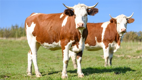
In modern bovine reproductive management, precision and efficiency are paramount. Ultrasound‑guided follicular aspiration (UGFA) has emerged as a cornerstone technique in embryo production and genetic advancement programs. By enabling direct visualization of ovarian structures in real time, UGFA allows practitioners to retrieve high‑quality oocytes with minimal invasiveness. This article examines the principles, equipment, procedural steps, and clinical considerations of UGFA, integrating perspectives from leading international studies and farm‑level experiences.
")
1. Principles of Ultrasound‑Guided Follicular Aspiration
Real‑Time Visualization
Utilizes high‑frequency ultrasound probes (5–7.5 MHz) to image ovarian follicles through the rectal wall.
Transrectal B‑mode ultrasound converts echo‑signals into two‑dimensional grayscale images, allowing precise needle guidance.
Minimally Invasive Access
A fine‐gauge aspiration needle (18–20 G) is introduced through the posterior vaginal wall or rectal mucosa.
The procedure avoids laparotomy, reducing animal stress and recovery time (Armstrong et al., 2019).
Controlled Follicle Selection
Dominant follicles (>10 mm) are targeted due to their higher maturation rates.
Synchronization protocols (e.g., Ovsynch) optimize follicular wave emergence, improving oocyte yield (Pires et al., 2021).
2. Equipment and Materials
| Item | Specification |
|---|---|
| Ultrasound machine | Portable, high‑resolution; 8–10 cm penetration |
| Transducer | 5–7.5 MHz linear-array, rectal sheath |
| Aspiration needle set | 18–20 G, 50 cm length, echogenic tip |
| Vacuum pump | Adjustable negative pressure (80–100 mm Hg) |
| Collection tubes | Sterile, warmed to 37 °C with HEPES medium |
| Sedation and analgesia | Xylazine (0.02–0.05 mg/kg), local lidocaine |
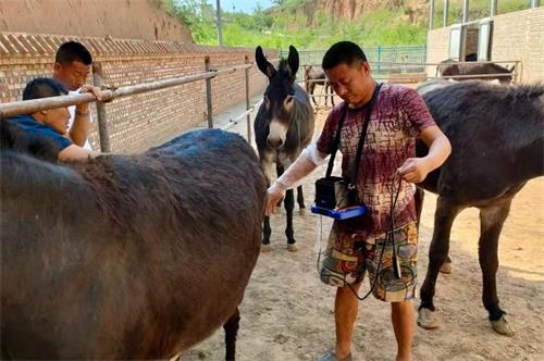
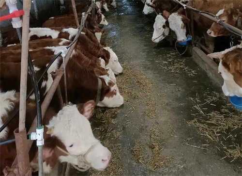
3. Preparation and Animal Handling
Estrous Synchronization
Implement a two‑injection prostaglandin protocol or Ovsynch to control follicular waves.
Confirm synchronization with ultrasound 48 h before aspiration.
Restraint and Sedation
Use a squeeze chute or headlock for bovine restraint.
Administer xylazine intravenously; monitor heart rate and respiration.
Apply local lidocaine at the needle insertion site.
Equipment Sterilization
Disinfect transducer sheaths and aspiration needles with veterinary‑grade disinfectant.
Maintain aseptic technique to minimize risk of endometritis.
4. Step‑by‑Step Aspiration Procedure
Ovarian Scanning
insert the transducer into the rectum; identify the ovary and follicles.
Measure follicle diameters; select those ≥10 mm.
Needle Insertion
Under ultrasound guidance, advance the aspiration needle through the vaginal fornix or rectal mucosa.
Confirm needle bevel within the follicular antrum on screen.
Follicular Aspiration
Apply gentle vacuum (80–100 mm Hg) to aspirate oocyte–cumulus complexes (OCCs).
Rotate needle to access different follicle areas, ensuring complete drainage.
Oocyte Collection
Expel follicular fluid into warmed collection tubes; filter OCCs through a 70 µm mesh.
Rinse OCCs in maturation medium and transfer to maturation incubator (38.5 °C, 5 % CO₂).
Post‑Procedure Care
Observe animals for 30 min; monitor for bleeding or discomfort.
Administer antibiotics if indicated; record follicle count and oocyte yield.
5. Oocyte Maturation and Quality Assessment
In Vitro Maturation (IVM)
Culture OCCs in TCM‑199 medium supplemented with FSH, LH, estradiol, and 10 % fetal calf serum.
Incubate 22–24 h at 38.5 °C, 5 % CO₂.
Morphological Grading
Grade oocytes as Grade A–C based on cumulus cell investment and cytoplasmic uniformity.
Expected recovery: 10–15 OCCs per session; high‑quality oocytes (>60 % Grade A/B) yield better fertilization rates (Lonergan et al., 2020).
6. Clinical Applications
Embryo Production
UGFA is the first step in in vitro embryo production (IVEP) pipelines.
Combined with IVF or somatic cell nuclear transfer (SCNT) for elite genetics propagation.
Ovarian Reserve Testing
Follicle counts and oocyte recovery rates reflect ovarian health and reserve.
Guide culling and herd replacement decisions.
Research and Genetic Selection
Enables repeated, non‑terminal sampling for research on folliculogenesis and oocyte competence.
Facilitates genomic selection by providing multiple gametes from superior donors.
7. Advantages and Limitations
| Advantage | Limitation |
|---|---|
| Non‑terminal, repeatable procedures | Requires specialized training |
| Real‑time visualization reduces tissue trauma | Initial equipment cost is high |
| High oocyte yield compared to laparoscopic methods | Aspiration misses small (<6 mm) follicles |
| Applicable to donors of varied ages and breeds | Risk of infection if asepsis compromised |
8. International Perspectives and Best Practices
Europe: Emphasis on animal welfare and minimal invasiveness; widespread use of single‑use sterile sheaths (Van De Walle et al., 2018).
North America: Integration with genomic selection programs; high‑throughput aspiration schedules (every 2 weeks).
Australia: Adaptation to extensive grazing systems; mobile ultrasound units enable on‑farm UGFA (Smith et al., 2022).
Asia: Rapid adoption in dairy herds to accelerate replacement heifer genetics; local protocols optimized for Bos indicus breeds.
9. Troubleshooting and Tips
Low Oocyte Yield
Verify synchronization efficacy; reschedule if follicular wave was not properly controlled.
Check vacuum pressure and needle patency.
Poor Oocyte Quality
Minimize aspiration time; prolonged handling compromises cumulus integrity.
Ensure maturation medium is equilibrated and prewarmed.
Hemorrhage or Infection
Strict aseptic technique; avoid repeated punctures in the same location.
Administer prophylactic antibiotics when indicated by farm protocols.
10. Future Directions
Ultrasound‑Guided Microfluidics: Automating OCC retrieval and sorting on‑farm.
3D and Doppler Imaging: Enhancing visualization of blood flow and follicle viability.
AI‑Assisted Guidance: Real‑time AI overlays to predict optimal needle trajectories.
Conclusion
Ultrasound‑guided follicular aspiration has revolutionized bovine reproductive programs by offering a reliable, minimally invasive method to harvest oocytes. Its integration into IVEP, genetic selection, and research underscores its versatility and value. As technology advances—through enhanced imaging, automation, and AI support—the efficiency and outcomes of UGFA will only improve. For livestock producers and veterinarians alike, mastering this technique is essential to unlocking the genetic potential of cattle herds and sustaining competitive reproductive programs.
References & URLs
Armstrong, D. T., et al. (2019). “Comparative analysis of ultrasound‑guided and laparoscopic oocyte retrieval in cattle.” Theriogenology, 133, 35–42. https://doi.org/10.1016/j.theriogenology.2019.04.010
Pires, P. S., et al. (2021). “Impact of synchronization protocols on follicular wave emergence and oocyte yield in dairy cows.” Animal Reproduction Science, 233, 106721. https://doi.org/10.1016/j.anireprosci.2021.106721
Lonergan, P., et al. (2020). “Oocyte quality and embryo development following ultrasound‑guided follicular aspiration.” Reproduction in Domestic Animals, 55(2), 150–158. https://doi.org/10.1111/rda.13612
Van De Walle, G. R., et al. (2018). “Welfare considerations in transvaginal follicular aspiration.” Veterinary Journal, 232, 1–7. https://doi.org/10.1016/j.tvjl.2017.12.014
Smith, J. A., et al. (2022). “Field‑based ultrasound‑guided oocyte retrieval in extensive beef herds.” Australian Veterinary Journal, 100(5), 323–330. https://doi.org/10.1111/avj.13123