As a livestock producer, accurately timing the breeding of cattle is one of the most critical factors influencing reproductive success, herd fertility rates, and overall farm profitability. In the era of precision livestock farming, non-invasive tools such as ultrasonography have become indispensable for monitoring reproductive physiology—especially follicular development—in cows. Among various ultrasonographic applications, evaluating ovarian follicles allows farmers and veterinarians to optimize breeding decisions, whether using natural service or artificial insemination (AI). In this article, I will explore how we use animal ultrasound scanners to evaluate follicular development in cattle, why this technology has become standard in modern cattle reproduction management, and what science tells us about its accuracy and benefits.
")
Understanding Follicular Development in Cattle
Follicular development is a highly dynamic, hormone-regulated process occurring in the ovaries of female cattle. Each estrous cycle includes several waves of follicular growth and regression, eventually leading to ovulation when conditions are optimal. Correctly identifying the stage of follicular development is critical for successful insemination. Misjudging the timing often results in poor conception rates and extended calving intervals—costly consequences for any farm.
Ovarian follicles develop in waves, typically two or three per cycle, with one dominant follicle eventually reaching ovulation while subordinate follicles regress. The precise growth pattern is influenced by multiple hormones, primarily follicle-stimulating hormone (FSH), luteinizing hormone (LH), and progesterone.
Early Follicular Phase: Multiple small antral follicles (2-5 mm in diameter) begin to grow under FSH stimulation.
Dominance Selection: One follicle becomes dominant, suppressing the growth of others through endocrine feedback.
Pre-Ovulatory Stage: The dominant follicle reaches 12–20 mm in size before ovulation.
Luteal Phase: After ovulation, the ruptured follicle transforms into a corpus luteum (CL), which secretes progesterone to maintain pregnancy or regulate subsequent cycles if fertilization does not occur.
By understanding these physiological stages, farmers and veterinarians can better assess when a cow is most fertile and time insemination with high accuracy.
Why Ultrasound is the Gold Standard for Follicle Monitoring
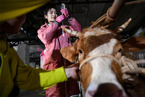
Traditional methods for estrus detection—such as behavioral observation, rectal palpation, or hormonal assays—are still widely used but have limitations in accuracy, labor intensity, and time requirements. In contrast, ultrasonography provides real-time, visual, and quantifiable information about ovarian structures.
Using a veterinary B-mode ultrasound scanner, such as the BXL-V50, allows us to clearly visualize:
Size and number of follicles
Shape and echogenicity of the dominant follicle
Presence and size of corpus luteum
Uterine tone and fluid content, offering indirect signs of estrus or pregnancy
This non-invasive technology dramatically reduces guesswork. In practical terms, I can scan my cows quickly and painlessly, often in under two minutes per animal, while obtaining highly reliable reproductive data. No sedatives or risky invasive procedures are required, and cows experience minimal stress.
")
How Follicle Evaluation Works in Practice
Daily Routine on the Farm
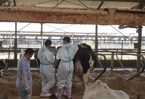
On my farm, we routinely conduct ovarian ultrasonography during the breeding season. Using a portable animal ultrasound scanner equipped with a rectal linear transducer, we insert the probe into the rectum to image the reproductive organs through the rectal wall. This method provides excellent image resolution of both ovaries and the uterus.
The following are typical observations:
Small follicles (3-5 mm): Early-stage wave follicles
Medium follicles (6-10 mm): Subordinate follicles still competing for dominance
Large follicles (12-20 mm): Dominant, pre-ovulatory follicles approaching readiness for breeding
With these measurements, we synchronize breeding plans based on the size and stage of follicular development, ensuring semen is delivered at the optimal time for fertilization.
Synchronization and Timed AI
Many commercial cattle operations utilize synchronization protocols such as Ovsynch or CIDR-based programs. Ultrasound allows us to verify whether these hormonal treatments are producing the intended ovarian responses:
Has the follicle reached sufficient size?
Is the corpus luteum regressing on schedule?
Is uterine tone consistent with estrus?
When ultrasound reveals a dominant follicle exceeding 15 mm with decreasing uterine tone, it is often the perfect moment to proceed with insemination.
Real-Life Example
Last spring, one of my high-value Holstein cows was synchronized for AI. Using ultrasound, I detected a 17 mm dominant follicle with soft uterine tone 36 hours after prostaglandin administration. We inseminated within 12 hours of this scan, and pregnancy was confirmed 30 days later. Without ultrasound, we might have either missed the window or inseminated too early, both of which would reduce pregnancy rates.
Scientific Validation of Ultrasound in Follicular Monitoring
Ultrasound's effectiveness for follicular evaluation is strongly supported by scientific research worldwide. For instance:
Pierson and Ginther (1998) demonstrated that transrectal ultrasonography accurately identified dominant follicle size and number, predicting ovulation within a narrow window of 24–48 hours.
Vasconcelos et al. (2001) confirmed that follicular measurements correlated strongly with conception success during timed AI protocols.
Lucy (2007) noted that ultrasound improved pregnancy rates by 10-20% compared to relying solely on behavioral estrus detection.
These studies reinforce what I see daily on my farm: data-driven breeding decisions result in higher conception rates and fewer repeat services.
")
The Advantages of Ultrasound in Modern Reproductive Management
From both a veterinary and farmer's perspective, ultrasound offers numerous advantages in reproductive management:
Non-Invasive and Low Stress: No sedation or discomfort for the animal.
Immediate Feedback: Real-time visualization allows for instant decision-making.
Accurate Follicle Measurement: Provides exact diameter of follicles, superior to rectal palpation.
Corpus Luteum Evaluation: Helps determine the effectiveness of synchronization protocols.
Reduced Open Days: Shortening the calving interval significantly boosts profitability.
On large commercial dairies or beef operations, ultrasound use translates into thousands of dollars saved annually through improved reproductive performance, optimized calving intervals, and better resource allocation.
Common Challenges and How to Overcome Them
While ultrasound is highly effective, several challenges exist:
Operator Training: Proficiency requires practice; recognizing follicles versus cysts or early CL can be challenging for beginners.
Equipment Cost: Although prices have dropped, quality ultrasound scanners remain a significant investment. However, the long-term benefits often outweigh initial costs.
Access to Animals: Handling facilities must allow safe and easy access for scanning without causing unnecessary stress or injury.
Investing in training and proper handling infrastructure has made these challenges very manageable on my farm. Over time, the skill becomes second nature.
Comparing Ultrasound to Hormonal Blood Testing
Some farms use blood tests to measure progesterone or estradiol levels for breeding timing. While hormonal assays offer useful biochemical information, they have limitations compared to ultrasound:
| Feature | Hormonal Testing | Ultrasound Scanning |
|---|---|---|
| Result Speed | Hours to days | Immediate |
| Follicle Size Info | Indirect | Direct Measurement |
| Corpus Luteum Data | Indirect | Direct Visualization |
| Uterine Condition | No info | Full Visualization |
| Cost Per Test | Repeated expense | Minimal after purchase |
In my experience, ultrasound offers a more comprehensive, immediate, and actionable set of data points for breeding management.
Global Adoption of Ultrasound in Cattle Reproduction
In the United States, Canada, Australia, and many parts of Europe, the use of ultrasound for reproductive monitoring has become nearly standard practice in progressive cattle operations. Several universities now offer short courses to train farmers and veterinary technicians in basic bovine ultrasonography.
For example, the Beef Cattle Institute at Kansas State University provides extensive training modules online, recognizing how important this skill has become in both dairy and beef industries (Beef Cattle Institute, 2023).
Additionally, precision farming technologies are integrating ultrasound data into herd management software, allowing farmers to track reproductive histories, follicular growth patterns, and pregnancy confirmations with unprecedented precision.
")
BXL-V50 Animal Ultrasound Scanner
Future Developments: AI Integration and Predictive Analytics
The future of follicular ultrasound monitoring may soon involve artificial intelligence (AI). Already, some research labs are developing algorithms that automatically identify follicles, measure their dimensions, and suggest optimal insemination windows based on real-time data patterns.
This could further enhance consistency and reduce the learning curve for new users. Combining AI-assisted ultrasound with wearable estrus monitors or automated hormone sampling may soon create highly accurate, integrated reproductive management systems.
Conclusion
For any cattle operation aiming to maximize reproductive success and profitability, accurate assessment of follicular development is essential. Animal ultrasound scanners provide real-time, reliable, and non-invasive data that allows farmers and veterinarians to optimize breeding timing with unmatched precision.
From my own farm experience and extensive research, it is clear that ultrasonography not only improves conception rates but also reduces costs associated with missed heats, repeated inseminations, and prolonged calving intervals. As technology advances, and as more producers worldwide embrace data-driven decision-making, ultrasound-guided reproductive management will continue to play a pivotal role in modern cattle breeding.
References
Pierson, R. A., & Ginther, O. J. (1998). Ultrasonography of the bovine ovary. Theriogenology, 29(1), 21–37.
Vasconcelos, J. L. M., et al. (2001). Timing of insemination and fertility in dairy cows following a synchronized ovulation regimen. Journal of Dairy Science, 84(2), 432–441.
Lucy, M. C. (2007). Reproductive loss in high-producing dairy cattle: where will it end? Journal of Dairy Science, 84(6), 1277–1293.
Beef Cattle Institute. (2023). Using Ultrasound to Evaluate Reproductive Status of Cattle. https://www.beefcattleinstitute.org/ultrasound-reproduction