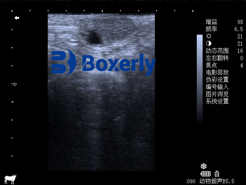
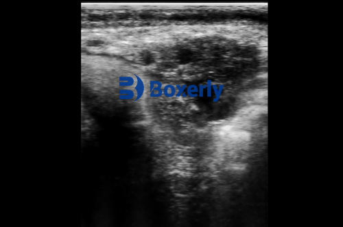
In modern cattle breeding, the practice of embryo transfer has become a vital strategy for rapidly multiplying the genetics of high-performing cows. This reproductive biotechnology allows breeders to collect fertilized embryos from elite donor cows and implant them into recipient cows, effectively enabling one superior cow to produce numerous offspring each year. However, the success of this process hinges on one critical foundation: the careful selection of both donor and recipient cows. Among the tools available to veterinarians and breeders, ultrasound imaging has emerged as an indispensable technology for pre-transfer screening. By offering clear insights into reproductive health and pregnancy status, ultrasound dramatically increases the efficiency and success rates of embryo transfer programs.

In this article, we will explore how ultrasound plays a pivotal role in donor and recipient selection, focusing on two primary areas: pregnancy diagnosis before embryo collection or transfer, and assessment of reproductive tract health to eliminate unsuitable animals. We'll also draw parallels with global practices and provide evidence from international veterinary standards.
Using Ultrasound to Detect Pregnancy Before Embryo Collection or Transfer
Embryo transfer protocols require cows to be in optimal reproductive condition and free of any existing pregnancies. A fundamental error in identifying a pregnant cow as a donor or recipient can lead to major reproductive failures, increased stress on the animal, and significant economic losses. This is especially crucial for recipient cows, who are expected to carry embryos to term after transfer.
In traditional practices, pregnancy detection was done using manual rectal palpation—a technique that, while effective in skilled hands, lacks the clarity and specificity of modern imaging. Ultrasound has revolutionized this process by providing real-time, high-resolution imaging of the uterus and ovaries. With a transrectal probe, veterinarians can accurately determine whether a cow is pregnant as early as 25 to 30 days post-breeding.
In donor cows, early pregnancy detection allows for the timely exclusion of unsuitable animals from the embryo flushing schedule, preventing unnecessary hormonal stimulation or surgical collection attempts. In recipients, this screening ensures that only cows capable of accepting and carrying embryos are selected.
Studies in the United States and Europe have demonstrated that farms utilizing ultrasound for pregnancy diagnosis prior to embryo transfer experience fewer failed implantations and miscarriages, leading to more predictable calving schedules and healthier offspring. This aligns with global recommendations from the International Embryo Technology Society (IETS), which advocates ultrasound as the preferred method for pregnancy confirmation prior to transfer.
Evaluating Reproductive Health with Ultrasound
A cow may not be visibly pregnant yet still be unsuitable as a donor or recipient due to hidden reproductive issues. In fact, subclinical reproductive tract diseases are common culprits behind failed embryo transfer attempts. Conditions such as endometritis (inflammation of the uterine lining), pyometra (uterine infection with pus accumulation), hydrosalpinx (fluid in the fallopian tubes), and various ovarian abnormalities may go undetected without advanced imaging.
This is where ultrasound proves invaluable. With a linear array transrectal probe, veterinarians can examine the structural integrity and physiological state of the uterus and ovaries. For example:
Endometritis appears as thickened uterine walls with fluid echoes.
Pyometra shows echogenic pus within the uterus.
Ovarian cysts and inactive ovaries can be visualized based on their size, shape, and internal consistency.
By identifying such abnormalities, breeders can remove unsuitable cows from the program, initiate appropriate treatments, or, in cases of poor prognosis, cull the animals early to reduce long-term losses.
International veterinary standards, such as those published in the Journal of Veterinary Reproductive Science and widely practiced in regions like Canada, Australia, and Germany, support the routine use of ultrasound as a first-line diagnostic tool in reproductive management. This proactive approach enhances embryo transfer success rates by ensuring only healthy, reproductively sound cows are used.

Case Studies and Global Perspective
In North America, large-scale cattle operations routinely employ ultrasound screening as part of their embryo transfer pre-check protocol. A 2022 report from the Beef Cattle Institute at Kansas State University found that farms implementing ultrasound-based screening prior to transfer saw up to a 15–20% improvement in conception rates compared to those relying on traditional methods.
Similarly, in New Zealand’s dairy sector, veterinarians report that ultrasound allows for precise estrous synchronization timing and better embryo survival. Farms using this technology also reported lower usage of hormonal drugs, reducing both cost and the risk of hormonal resistance.
These findings are not isolated. A comparative study conducted in Germany by Reproduction in Domestic Animals (2021) highlighted that ultrasound-screened recipients were 30% less likely to reject embryos due to underlying reproductive issues that were pre-emptively identified and treated.
Practical Application: Step-by-Step Ultrasound Use in Embryo Transfer Programs
Initial Health Assessment
All cows undergo a general health check to rule out visible illness or systemic issues.Ultrasound Pregnancy Diagnosis
Using a transrectal linear probe, the veterinarian checks for early pregnancy. This ensures:Donors are not already carrying embryos
Recipients are ready to receive embryos
Reproductive Tract Scanning
Evaluate the uterus, cervix, fallopian tubes, and ovaries. Identify any signs of:Uterine infections or fluid accumulation
Ovarian inactivity or cysts
Structural abnormalities
Classification and Selection
Based on the scan, cows are classified as:Fit for embryo work
Require medical treatment
Recommended for culling
Follow-up Monitoring
Post-treatment cows may undergo re-scanning to assess recovery before reintegration into the program.

Beyond Cattle: Insights from Aquaculture Ultrasound Practices
Interestingly, the same principle applies in other animal sectors. In aquaculture, for instance, ultrasound is commonly used to evaluate the reproductive condition of fish like the black rockfish (Sebastes schlegelii). Researchers use ultrasound to assess gonad maturity, which helps optimize breeding programs. This cross-species application underscores the versatility of ultrasound in animal reproductive management.
Conclusion
Ultrasound technology is more than just a diagnostic tool—it is a strategic asset in reproductive management, especially for embryo transfer in cattle. By identifying existing pregnancies and reproductive tract diseases early, ultrasound helps farmers and veterinarians select the right animals, saving time, money, and effort.
Globally, the trend is clear: farms that integrate ultrasound into their reproductive protocols consistently achieve higher pregnancy rates, healthier calves, and better genetic outcomes. For any breeder aiming to maximize embryo transfer success, investing in routine ultrasound screening is not just advisable—it is essential.
Reference Sources:
International Embryo Technology Society (IETS). (2023). Embryo Transfer Guidelines. https://www.iets.org
Whitaker, D. A., & Smith, E. (2021). Veterinary Ultrasonography in Food-Producing Animals. Journal of Veterinary Imaging.
Beef Cattle Institute. (2022). “Use of Ultrasound for Growth Evaluation in Cattle.” https://www.beefcattleinstitute.org/ultrasound-growth
Reproduction in Domestic Animals. (2021). “Success Factors in Embryo Transfer Programs”. Wiley Online Library.


