In modern swine production, precision in reproductive monitoring is key to improving productivity and minimizing losses. As sow pregnancy is one of the most critical stages influencing piglet output and herd efficiency, producers around the world have turned to Veterinary ultrasound as an essential, non-invasive, and real-time diagnostic tool. By using ultrasound to monitor both the presence and number of fetuses and to plan breeding or farrowing schedules, producers can significantly improve outcomes in sow reproduction. In this article, we’ll explore how ultrasound is applied to count fetuses, determine gestation timelines, and discuss why this practice is becoming standard in progressive swine operations globally.

Ultrasound Monitoring of Sow Pregnancy: Practical Methods in the Field
Timing of Scanning
In swine herds, timing is everything. The ideal window for determining pregnancy and fetal count via ultrasound is typically around day 25 to day 30 after mating. Conducting two separate ultrasound sessions within this time frame allows farmers to estimate litter size more accurately and detect potential reproductive failures early, enabling timely rebreeding decisions.
Foreign practitioners commonly follow this dual-scan approach as well, acknowledging that single checks can lead to miscounts due to embryonic resorption or scanning too early. A second scan improves the reliability of fetal detection, especially for gilts or sows with a history of low litter size.
Positioning and Handling During Ultrasound Scanning
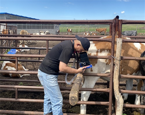
Contrary to what some might expect, sows do not need to be forcefully restrained during external abdominal scanning. Most remain calm if handled gently. The preferred posture is lateral recumbency (lying on the side), but scanning can also be performed when the sow is standing, feeding, or even resting in a prone position. If the sow is aggressive or hard to reach, handlers may use gates, boards, or gentle tools like a pig catcher to safely position the animal in a corner.
In modern, large-scale pig operations, the scanning is often performed directly in farrowing crates or individual pens, minimizing animal movement. When using the transrectal approach—less common in pigs but occasionally necessary for early pregnancy checks or specific research—the sow must be properly restrained in a standing position.
Optimal Scanning Locations and Probe Orientation
The external probe is typically placed on the lower abdominal region near the last pair of mammary glands, just above the udder. This area provides the most direct access to the uterus in early pregnancy. As gestation progresses, the uterus enlarges and extends forward under the ribs, requiring the operator to adjust the scanning site accordingly.
Unlike in cattle or sheep, the sparse hair on pigs means shaving is generally not required before scanning. However, it is essential to clean the area, removing any mud or debris. Coupling gel should be liberally applied to ensure effective transmission of ultrasound waves.
For internal transrectal exams, which are rare but used in research or reproductive studies, disinfectant or water is typically used to moisten the probe, avoiding the need for gel and ensuring hygiene during insertion.
Scanning Techniques: Count Estimation and Fetal Confirmation
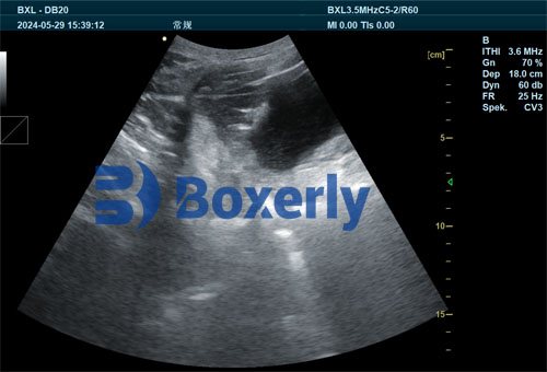
In early pregnancy (around day 25–30), the embryos are still small, making them harder to detect with speed. For accurate fetal count estimation, the probe should be held firmly against the abdominal wall. The scanning angle is important: point the probe towards the pelvic cavity or at a 45-degree angle toward the opposite upper abdomen.
Operators perform a slow, fan-like sweep in both horizontal and vertical directions. This technique helps identify individual fluid-filled gestational sacs. Quick sliding motions across the skin should be avoided, as they reduce visibility of early embryonic structures. Sometimes gentle inward pressure is applied to push aside intestinal loops and bring the uterus closer to the probe.
It’s customary to start with the right side of the sow, systematically scanning for each visible gestational sac. After recording the count on one side, the operator repeats the same process on the left side. Adding both counts gives an approximate total number of fetuses, which can later guide feeding, management, and culling decisions.
Why Foreign Producers Rely on Ultrasound Technology
In the United States, Denmark, Germany, and other leading pork-producing countries, ultrasound pregnancy checks have become a standard management practice. International swine veterinarians and technicians emphasize its core advantages:
Non-invasive and Safe: No stress or physical trauma to the sow.
Accurate Estimations: Fetal count and pregnancy status are determined with over 90% accuracy.
Efficient Labor Use: Ultrasound scanning can be done quickly and repeatedly without moving the sow from her pen.
Early Decision-Making: Sows not pregnant by day 30 can be promptly re-entered into the breeding cycle.
In high-performing operations, ultrasound also supports gilt selection by confirming early fertility performance and ensuring non-pregnant females don’t occupy valuable resources unnecessarily.
Scheduling and Reproductive Planning Based on Scan Results
Once pregnancy is confirmed and fetal counts are estimated, the sow’s future management can be more precisely structured. For instance:
Feeding Adjustments: The number of fetuses directly influences feed energy requirements, especially in late gestation.
Farrowing Calendar: Ultrasound can help estimate gestation progression and expected farrowing windows, improving farrowing crate utilization.
Culling Strategy: Sows with consistently low fetal counts or reproductive failure can be identified and replaced, boosting overall herd productivity.
Addressing Challenges: Operator Training and Interpretation
Although ultrasound is highly effective, it is operator-dependent. International literature emphasizes that training is essential. Misinterpretation of images—such as mistaking fluid-filled intestines for gestational sacs—can lead to false positives or inaccurate fetal counts.
Advanced devices with image enhancement and Doppler functionality help reduce user error by offering clearer contrast between tissues. Some models even have fetal heart rate monitoring to further confirm viability.
Training programs in countries like the Netherlands, Canada, and Australia have included ultrasound interpretation as part of standard pig veterinary education. The goal is to ensure every operator can read the B-mode images effectively, enhancing reliability across different herd conditions.
Broader Application in Veterinary Practice: From Sows to Fish
Interestingly, the concept of using ultrasound to monitor reproductive status is not limited to pigs. In aquaculture, species such as Sebastes schlegelii—a type of marine rockfish—are also examined using ultrasound to determine gonadal maturity and reproductive output. This cross-species application underscores ultrasound’s growing relevance in all areas of animal reproduction management.
Future Outlook: Data-Driven Swine Farming
As precision livestock farming becomes the new standard, integrating ultrasound data into digital herd management systems is the next logical step. Several foreign companies now offer ultrasound machines with USB export, Bluetooth syncing, or cloud platforms. These features allow real-time sharing of scan results with veterinarians and farm managers, facilitating faster and more coordinated decisions.
Combining fetal count data with other metrics such as sow body condition, backfat thickness, and parity records enables a more holistic reproductive strategy—one that improves sow longevity, enhances piglet survival, and reduces unnecessary costs.
Conclusion
Ultrasound scanning has revolutionized sow reproductive management across the world. By providing reliable, real-time insight into pregnancy status and fetal number, it supports better breeding decisions, optimized feeding, and enhanced economic outcomes. On both small family farms and large-scale commercial operations, this technology has proven itself indispensable.
From pig barns in Iowa to tech-integrated farms in Europe and Asia, ultrasound is not just a diagnostic tool—it’s a decision-making partner in modern swine production.
References:
Whitaker, D. A., & Smith, E. (2021). Veterinary Ultrasonography in Food-Producing Animals. Journal of Veterinary Imaging.
Van Nes, A., & Loos, C. (2023). Ultrasound Applications in Swine Herd Reproductive Management. Swine Health Professionals International.
https://www.beefcattleinstitute.org/ultrasound-growth

