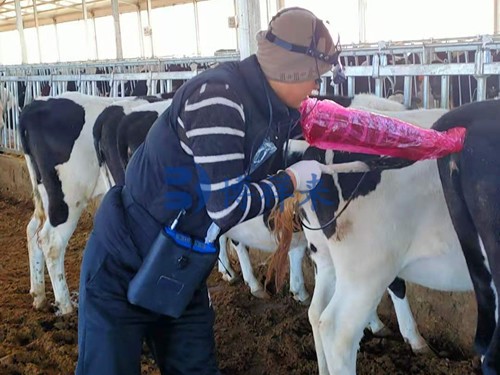
For modern livestock producers and veterinarians, timely diagnosis and effective management of reproductive health are essential to maintaining productivity, particularly in dairy and breeding animals. Among the many diagnostic tools used in reproductive management, veterinary ultrasonography (ultrasound) has emerged as one of the most reliable, non-invasive, and real-time methods for evaluating uterine conditions and monitoring uterine involution after parturition. In this article, we explore how ultrasound helps in diagnosing uterine diseases and in tracking uterine recovery postpartum—topics that have gained increasing importance in veterinary medicine globally.
")
Diagnosing Uterine Diseases Using Ultrasound
Uterine diseases such as endometritis, pyometra, and uterine tumors are significant reproductive health issues that can lead to infertility, decreased milk production, and extended calving intervals in dairy cows and other farm animals. Early and accurate diagnosis is critical for implementing appropriate treatments and minimizing economic losses.
Traditional diagnostic methods, such as rectal palpation and vaginal examination, have limitations—especially when the disease is in the early stages or when subtle structural abnormalities exist. Here, ultrasound imaging provides a clearer, more objective window into the uterine environment.
Common Ultrasonographic Signs of Uterine Disorders:
Chronic Endometritis: On ultrasound, the uterine cavity often appears expanded with indistinct margins and floating echogenic material, indicating inflammatory exudate. The fluid may be accompanied by snowflake-like particles, suggesting infection.
Pyometra: A condition marked by the accumulation of pus in the uterine lumen. Ultrasound typically reveals an enlarged uterine body, increased and clarified wall echogenicity, and the presence of hypoechoic fluid regions. It's important to differentiate this from early pregnancy, as both conditions can display uterine fluid pockets.
Uterine Tumors: Although rare in livestock, tumors such as adenocarcinomas or leiomyomas can sometimes be visualized as irregular, solid masses with variable echogenicity. Continuous monitoring and biopsy may be necessary for confirmation.
Ultrasound not only supports visual diagnosis but also enables real-time assessment of treatment efficacy. For instance, post-treatment scanning can confirm the resolution of uterine fluid or inflammation, helping avoid unnecessary further interventions.
Monitoring Uterine Recovery in Dairy Cows Postpartum
Uterine involution—the process by which the uterus returns to its pre-pregnant size and state—is essential for resuming normal reproductive cycles and ensuring successful rebreeding. Traditionally evaluated through rectal palpation, uterine recovery has been difficult to quantify due to its dependence on practitioner experience.
By contrast, ultrasound provides measurable, repeatable parameters that make uterine involution monitoring more standardized and accurate.
Benefits of Using Ultrasound for Postpartum Monitoring:
Objective Measurement: With ultrasound, one can measure the diameter of the cervix, uterine body, and horns, allowing for a more precise timeline of recovery.
Continuous Monitoring: Unlike single-point palpation, serial ultrasonography can track changes in uterine morphology over time, revealing normal versus abnormal patterns of involution.
Detection of Subclinical Disorders: Conditions like subclinical endometritis or retained fetal membranes might not be palpable but are often visible on ultrasound.
Key Findings from Research:
Recent studies suggest that the duration of uterine involution varies between primiparous (first-time) and multiparous cows. For example:
Primiparous cows generally complete uterine recovery within 40 days.
Multiparous cows may take up to 50 days to fully involute.
In one monitored case, the cervix diameter in multiparous cows measured 97.00 mm on day 1 postpartum, which reduced to 36.14 mm by day 27, reaching a stable condition. The uterine wall thickness showed a pattern of thickening and then thinning—starting at 7.00 mm on day 1, increasing to 19.67 mm on day 22, and decreasing again to 13.00 mm on day 27.
Such detailed measurements help veterinarians identify delayed uterine involution early and apply interventions like uterine massage or intrauterine medications. This ensures cows return to estrus sooner, ultimately shortening the calving interval—a critical economic factor in dairy operations.
")
International Perspectives and Applications
Globally, the use of ultrasound in veterinary reproductive health has grown significantly. In countries like the United States, Canada, and parts of Europe, portable Veterinary ultrasound devices are standard tools on dairy farms. These devices allow field veterinarians to carry out on-site diagnostics and offer evidence-based reproductive management.
Researchers in the Netherlands and Germany, for instance, have emphasized the role of ultrasonography in improving reproductive efficiency and animal welfare. American studies also highlight the integration of ultrasound with reproductive protocols such as Ovsynch, where determining uterine health status enhances timed artificial insemination success.
Additionally, ultrasound data are now being linked with herd management software to analyze reproductive performance trends, helping farmers make more informed decisions regarding culling, breeding, and veterinary interventions.
Future Directions in Uterine Monitoring
While ultrasonography has already improved the clinical management of uterine conditions, there are promising developments on the horizon:
Endoscopic-Ultrasound Integration: Researchers suggest that combining ultrasound with uterine endoscopy (hysteroscopy) could offer both visual and microscopic evaluations, especially for research on high-producing cows.
Molecular Imaging: Future studies may include cellular and molecular-level investigations into postpartum uterine recovery. Such work would enhance our understanding of hormonal, immune, and structural changes, potentially leading to targeted pharmaceutical treatments.
Automated Ultrasound Systems: Robotic or AI-assisted scanning systems are being developed to reduce user bias and allow for consistent, automated monitoring of uterine involution in large herds.
Practical Takeaways for Farmers and Veterinarians
Start Scanning Early: Begin ultrasound monitoring within the first week postpartum to establish a baseline for each cow’s uterine recovery.
Track Progress Weekly: Follow-up scans every 7–10 days can catch deviations from normal involution patterns.
Compare with Reproductive History: Past parturition issues may predispose cows to slower recovery, making them prime candidates for closer ultrasound monitoring.
Integrate with Herd Software: Link ultrasound findings with breeding and health records for better herd-level reproductive analysis.
Conclusion
Ultrasonography has revolutionized the diagnosis of uterine diseases and the monitoring of uterine involution in livestock. By offering real-time, non-invasive, and quantitative insights into uterine health, this technology has become a cornerstone of modern veterinary reproductive care. Whether it’s identifying early endometritis or tracking postpartum uterine recovery, ultrasound enables faster, more accurate decision-making—ultimately improving animal welfare, fertility outcomes, and farm profitability.
As more research emerges and tools become even more accessible, the role of ultrasound in veterinary practice will only grow. For dairy producers and veterinarians alike, embracing ultrasound technology is a step toward more precise, efficient, and compassionate animal care.
")
References
Sheldon, I. M., & Dobson, H. (2004). Postpartum uterine health in cattle. Animal Reproduction Science, 82–83, 295–306. https://doi.org/10.1016/j.anireprosci.2004.04.006
Morrow, D. A. (2018). Current Therapy in Theriogenology. Elsevier Health Sciences.
Whitaker, D. A., & Smith, E. (2021). Veterinary Ultrasonography in Food-Producing Animals. Journal of Veterinary Imaging.
Beef Cattle Institute. (2023). “Use of Ultrasound for Growth Evaluation in Cattle.” https://www.beefcattleinstitute.org/ultrasound-growth