Ultrasound has revolutionized veterinary diagnostics, providing non-invasive insights into animal health, particularly in reproductive management. In canine breeding, early pregnancy diagnosis via ultrasound has become standard practice among veterinarians and breeders. However, concerns have persisted regarding the potential risks ultrasound may pose to the developing embryos during early gestation. This article aims to explore the safety profile of ultrasound use in early canine pregnancy, integrating both scientific research and global veterinary perspectives.
")
Ultrasound in Canine Reproduction: A Global Veterinary Tool
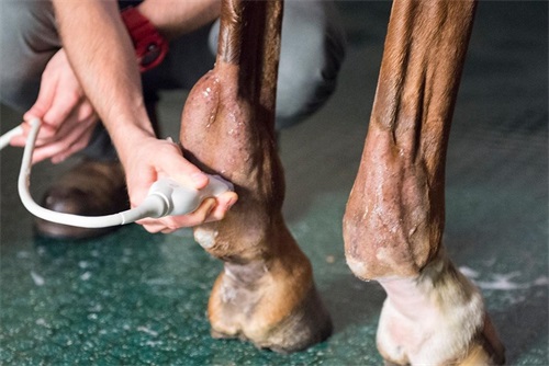
Across the world, veterinarians rely on B-mode ultrasound (brightness mode) to confirm pregnancy, determine fetal number, assess viability, and monitor development. In North America and Europe, the technique is widely accepted due to its safety and reliability. Veterinary guidelines—such as those published by the American College of Veterinary Radiology (ACVR) and the European Veterinary Diagnostic Imaging Society (EVDI)—recommend ultrasound as the preferred method for early pregnancy diagnosis in dogs, typically starting around day 25 post-ovulation.
One reason for its widespread use is the non-ionizing nature of ultrasound. Unlike X-rays, which emit harmful radiation, ultrasound uses high-frequency sound waves to create images, minimizing the risk of genetic or cellular damage.
Understanding the Safety Thresholds of Ultrasound Exposure
Scientific investigations have consistently shown that diagnostic ultrasound, when applied within recommended parameters, does not harm maternal or fetal health. A key consideration in determining safety is the intensity and duration of ultrasound exposure. Both factors are inversely related: the longer the examination, the lower the safe intensity must be to prevent biological effects.
For Veterinary ultrasound Equipment used in canine pregnancy checks, the emitted sound energy generally falls between 1% to 10% of the lowest recognized threshold for potential thermal damage. Moreover, the typical scanning duration for a pregnancy check is short—often under five minutes—far below the exposure time considered risky in thermal index (TI) or mechanical index (MI) safety models.
")
Thermal, Mechanical, and Cavitation Effects: Are They a Concern?
Despite the general consensus on safety, it is essential to understand the biological effects of ultrasound. These include:
Thermal Effect: Heat generation due to tissue absorption of sound energy. Excessive heating can theoretically harm embryonic tissue, particularly in early development stages.
Mechanical Effect: Also known as acoustic streaming or radiation force, it can affect cellular structures.
Cavitation: Formation of gas bubbles in tissues, which can collapse and cause cellular injury, although this is more of a concern with high-intensity therapeutic ultrasound, not diagnostic devices.
Fortunately, veterinary B-mode ultrasound is designed to avoid these risks. The FDA and international regulatory bodies have set limits for ultrasound output specifically to prevent such effects. When veterinarians use proper scanning techniques and avoid prolonged, localized exposure, the risk is considered negligible.
Empirical Studies: What the Data Shows
A number of experimental studies support the safety of ultrasound during canine pregnancy:
In a study involving 30 pregnant bitches scanned regularly during gestation days 57 to 64, there were no cases of abortion, dystocia, or stillbirth. All litters were born healthy, with no visible abnormalities or developmental defects.
Comparative trials involving rabbits and pigs subjected to similar diagnostic ultrasound showed no increase in embryonic loss or congenital anomalies.
In equine and ovine veterinary medicine, ultrasound is routinely used for pregnancy monitoring and has not been linked to fetal compromise.
These findings reflect a strong consensus: under routine clinical conditions, ultrasound poses no observable harm to either mother or offspring.
")
International Veterinary Consensus on Safe Practice
Veterinarians worldwide share a unified approach to safe ultrasound application in reproductive diagnostics. Key best practices include:
Minimizing scan time during early gestation (typically under 5 minutes).
Using low-output settings tailored to reproductive imaging.
Avoiding Doppler ultrasound during the first 30 days of pregnancy, as it produces higher acoustic outputs than B-mode imaging.
In countries like the United States, Canada, Germany, Australia, and Japan, veterinary schools and professional bodies emphasize these safety protocols during training and continuing education. As a result, ultrasound misuse is rare, and when properly applied, the benefits far outweigh any theoretical risks.
Practical Applications and Owner Considerations
For dog breeders and pet owners, the reassurance that ultrasound is safe makes it easier to plan for whelping, evaluate litter size, and manage high-risk pregnancies. Early diagnosis also allows for early nutritional and medical interventions, improving overall outcomes for both the bitch and the pups.
When owners ask whether ultrasound can harm unborn puppies, veterinarians can confidently point to decades of safe usage and a wealth of peer-reviewed evidence supporting its use.
")
Important Considerations for Ultrasound Timing
While safety is well established, timing remains important:
Before Day 21: It is difficult to detect viable pregnancies, and scanning too early may result in false negatives.
Day 25–30: This is the ideal window for accurate diagnosis, with visible gestational sacs and developing embryos.
After Day 40: Fetal structures become more defined, allowing assessment of viability, heartbeat, and growth parameters.
Doppler ultrasound, which visualizes blood flow, should ideally be used later in gestation when fetal organs are more developed. Even then, practitioners are advised to limit its use to short bursts.
Global Research Trends and Technological Advancements
In recent years, Veterinary ultrasound machines have become more sophisticated, with improved image resolution, portability, and automated measurements. Research into using 3D and 4D ultrasound for reproductive diagnostics is ongoing, particularly in advanced veterinary centers in Europe and Asia.
These advancements allow for more precise monitoring of fetal development and may one day help detect anomalies earlier than ever before—all while adhering to strict safety standards.
Conclusion
The global veterinary community strongly supports the use of ultrasound in early canine pregnancy as a safe, effective, and essential diagnostic tool. With appropriate training, calibrated equipment, and adherence to scanning protocols, ultrasound poses no significant risk to embryonic development.
From research in multiple species to decades of clinical experience, the evidence is overwhelmingly clear: diagnostic ultrasound, when used responsibly, is not only safe but indispensable in modern veterinary reproductive care. Breeders, pet owners, and veterinarians alike can rest assured that this technology plays a vital role in promoting the health and welfare of both pregnant dogs and their future litters.
link: https://www.bxlimage.com/nw/1208.html
tags: