Veterinary medicine has undergone a significant transformation with the growing adoption of ultrasound technology. Once considered a specialized tool limited to referral hospitals, ultrasound has now become a standard diagnostic modality across all types of veterinary practice—including rural farm settings and primary care facilities. From pregnancy checks in livestock to internal organ evaluation in companion animals, the utility and accessibility of ultrasound are expanding rapidly. This article explores how ultrasound supports accurate diagnoses, enhances treatment efficiency, and improves outcomes in animal care, especially for large-animal and farm-based veterinary practices.
")
Understanding the Role of Ultrasound in Veterinary Diagnostics
Ultrasound is a non-invasive, real-time imaging technique that uses high-frequency sound waves to generate visual representations of internal organs and structures. Unlike X-rays, which are better suited for visualizing bones, ultrasound excels in imaging soft tissues such as muscles, organs, and blood vessels. Its safety, portability, and repeatability make it particularly suitable for both routine and emergency veterinary diagnostics.
In veterinary settings, ultrasound is often used as a complementary tool to physical exams, laboratory tests, and radiography. It offers a detailed view of internal anatomy without requiring surgical intervention or, in many cases, even sedation. This is especially important in animals that may be stressed or at risk under anesthesia, such as pregnant livestock, neonatal animals, or patients with compromised health.
Diagnostic Applications Across Species
Reproductive Health and Pregnancy Monitoring
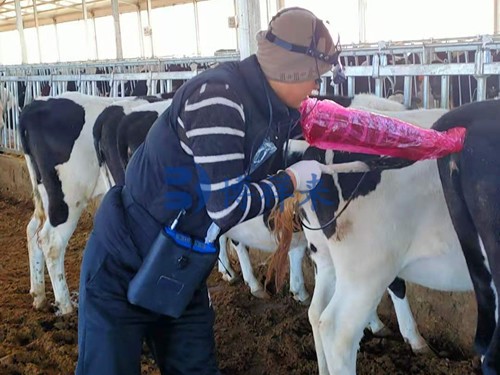
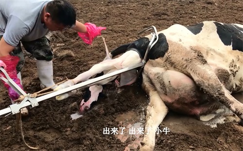
One of the most common applications of ultrasound in large-animal practice is reproductive management. In cattle and horses, veterinarians use ultrasound to:
Confirm pregnancy and estimate gestational age
Monitor fetal development and detect abnormalities
Evaluate ovarian structures and uterine health
Assist in breeding programs, such as embryo transfer and artificial insemination
For farm owners, timely and accurate pregnancy diagnosis is crucial for managing herd productivity and planning breeding schedules. portable ultrasound devices now allow veterinarians to conduct on-site pregnancy checks with minimal disruption to the animal’s routine.
Abdominal and Organ Evaluation
Abdominal ultrasound is widely used to assess the health of vital organs, including:
Liver: Detects inflammation, fatty infiltration, tumors, and biliary obstruction.
Kidneys: Identifies signs of infection, kidney stones, or structural abnormalities.
Bladder: Detects stones, thickening, or blockages.
Gastrointestinal Tract: Helps diagnose obstruction, foreign bodies, or motility disorders.
Pancreas and Adrenal Glands: Evaluates for enlargement, cysts, or tumors.
In ruminants and swine, ultrasound is particularly useful for diagnosing digestive issues, such as displaced abomasum or peritonitis, which may not be readily apparent through physical examination alone.
")
Cardiac and Thoracic Imaging
Thoracic ultrasound allows visualization of the heart and lungs, particularly useful in emergency situations or when animals present with respiratory distress. Focused cardiac ultrasound can help detect:
Pericardial effusion
Cardiac enlargement or failure
Pleural fluid
Pulmonary lesions
Especially in foals and calves, where congenital heart defects may arise, early identification via ultrasound can guide timely treatment or surgical intervention.
Musculoskeletal and Tendon Assessment
Musculoskeletal ultrasound is a valuable tool in equine and athletic livestock care. It allows real-time imaging of:
Tendons and ligaments
Joint effusions
Soft tissue injuries
Abscesses or hematomas
Veterinarians can monitor the healing process of injuries and adjust treatment protocols accordingly, helping animals recover faster and reducing downtime.
Ocular and Neurological Diagnostics
Ocular ultrasound provides insight into eye health when direct visualization is limited due to trauma or opacity. It can reveal:
Retinal detachment
Lens luxation
Vitreal hemorrhage
Intraocular tumors
Ultrasound of the neck can also assist in identifying masses, thyroid or salivary gland abnormalities, lymph node enlargement, or foreign bodies.
")
Operational Advantages for Veterinary Practices
Faster Diagnosis, Better Treatment Outcomes
One of the most significant benefits of ultrasound is its ability to provide real-time information. In urgent cases, such as internal bleeding or colic, ultrasound can dramatically shorten the time to diagnosis and intervention. This not only improves survival rates but also reduces the need for more invasive and costly procedures.
Portability and Field Readiness
Modern Veterinary ultrasound machines are lightweight and battery-operated, allowing veterinarians to carry them into barns, pastures, or remote farm locations. This is especially beneficial in areas with limited access to veterinary hospitals or where transporting large animals is impractical.
Reduced Need for Referrals
For many routine diagnostic scenarios, in-house or on-site ultrasound eliminates the need to refer animals to specialized centers. This reduces wait times, lowers costs, and keeps diagnostic and treatment decisions in the hands of the attending veterinarian.
Cost-Effectiveness for Farmers
Early and accurate diagnosis leads to better treatment outcomes and avoids losses due to misdiagnosis or delayed care. For example, detecting a uterine infection in a breeding cow early through ultrasound may prevent infertility and loss of production. For farm operations, this translates directly into increased profitability and herd efficiency.
")
Considerations When Implementing Ultrasound in Farm Practice
Training and Skill Development
While ultrasound is non-invasive and user-friendly, it still requires proper training to interpret images accurately. Many veterinary schools and continuing education programs now offer specialized training in ultrasound for large animals. Developing scanning proficiency is essential for maximizing the utility of the device.
Choosing the Right Equipment
Veterinarians should consider the following when selecting a machine:
Durability and portability for field use
Probe types and compatibility for different applications (linear for tendons, convex for abdominal scans, etc.)
Battery life and wireless capabilities
Image quality and real-time video recording
Ease of sterilization and maintenance
A flexible system that allows upgrades and probe changes can accommodate a wide variety of farm animal diagnostic needs.
")
Conclusion: Ultrasound as a Core Tool in Modern Veterinary Care
Ultrasound has become an indispensable diagnostic tool in modern veterinary medicine. For animal farm operators and livestock caretakers, it provides a window into the animal’s internal health—helping to make informed decisions quickly, safely, and cost-effectively.
Whether it’s checking pregnancies in cattle, identifying organ abnormalities in swine, or diagnosing tendon injuries in horses, veterinary ultrasound improves clinical outcomes and operational efficiency. As technology continues to evolve, the accessibility and impact of ultrasound on animal health will only grow, making it a worthy investment for any veterinary practice or farm operation.
References
DeFrancesco T, Royal K. A survey of point-of-care ultrasound use in veterinary general practice. Education in the Health Professions. 2018;1(2):50. https://doi.org/10.4103/ehp.ehp_21_18
Pelchat J, Chalhoub S, Boyson SR. The use of veterinary point-of-care ultrasound by veterinarians: A nationwide Canadian survey. Canadian Veterinary Journal. 2020. https://www.ncbi.nlm.nih.gov/pmc/articles/PMC7659883/
Lyssens A, Lekane M, Gommeren K, et al. Focused cardiac ultrasound to detect pre-capillary pulmonary hypertension. Front Vet Sci. 2022. https://doi.org/10.3389/fvets.2022.830275
Janson CO, Hezzell MJ, Oyama MA, et al. Focused cardiac ultrasound in emergency feline care. J Vet Emerg Crit Care. 2020. https://doi.org/10.1111/vec.12957
Assawarachan SN, Chuchalermporn P, Maneesaay P, et al. Evaluation of hepatobiliary ultrasound scores in dogs. Vet World. 2019. https://doi.org/10.14202/vetworld.2019.1266-1272
Kumar V, Kumar A, Varshney AC, et al. Diagnostic imaging of canine hepatobiliary affections: a review. Vet Med Int. 2012. https://doi.org/10.1155/2012/672107
Nussbaum LK, Scavelli TD, Scavelli DM, et al. Abdominal ultrasound in hyperthyroid cats. J Vet Intern Med. 2015. https://doi.org/10.1111/jvim.13369
O'Neill DG, Khoo JSP, Brodbelt DC, et al. Risk factors for hypothyroidism in dogs. Canine Med Genet. 2022. https://doi.org/10.1186/s40575-022-00123-8
Pollard RE, Bohannon LK, Feldman EC. Incidental thyroid nodules in dogs. Vet Radiol Ultrasound. 2014. https://doi.org/10.1111/vru.12181
Feliciano MAR, Uscategui RAR, Maronezi MC, et al. Ultrasonography in canine mammary tumors. PLoS One. 2017. https://doi.org/10.1371/journal.pone.0178143
Pagani E, Tursi M, Lorenzi C, et al. Ultrasonographic features of adrenal gland lesions in dogs. BMC Vet Res. 2016. https://doi.org/10.1186/s12917-016-0895-1