


BXL-S300
The BXL-S300 is an upgraded version of the S200, designed to deliver superior image clarity with its 128-element probe and true color Doppler imaging. This portable veterinary ultrasound system offers enhanced resolution for more precise diagnostics across a wide range of animals. With an intuitive button layout, the BXL-S300 ensures quick access to key functions, helping veterinarians save time during scanning. Whether used for pregnancy detection or reproductive management, this model provides reliable, high-definition images in real-time. Compact, efficient, and easy to operate, the BXL-S300 is built to perform in fast-paced clinical and farm environments.
Contact Us
Specifications
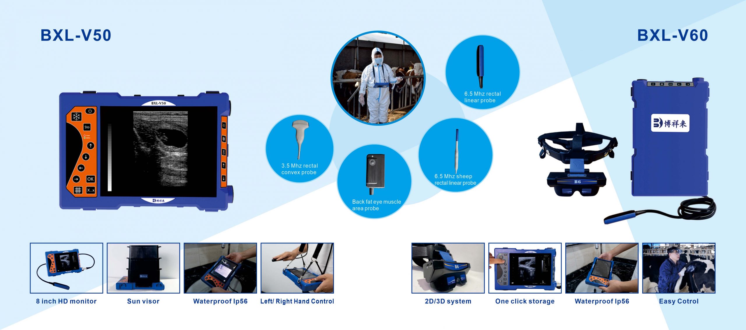
- IPX7 Waterproof &Dustproof
- Lightweight (400g with Probe)
- ≥6 Hours Battery Life
- 5G Wireless Transmission
- Doppler CF/PDI/DPDI Modes
- 128-element Rectal Probe
- ≥50 Veterinary Presets
- Tablet Display (Included)

Applications
The BXL-S300 Doppler Cattle & Horse Pregnancy Scanner revolutionizes large animal reproductive diagnostics with its IPX7 waterproof design and wireless imaging capabilities. Engineered for cattle, horses, and camelids, this 128-element rectal probe system delivers clinical-grade clarity in demanding farm environments.
- Advanced Doppler Imaging: Triple-synchronization mode (B+CF+PW) enables real-time blood flow monitoring for precise pregnancy confirmation, supported by 60fps frame rate for dynamic organ visualization.
- Military-Grade Portability: 400g ultra-lightweight body with 6-hour battery, IPX7 waterproofing, and belt-mount design withstands rigorous field use while enabling hands-free operation.
- AI-Powered Ranch Management: Auto-recognition of 50+ species-specific anatomies with intelligent backfat/eye muscle measurement, seamlessly integrated with electronic ear tags for automated reporting.
- 5G Wireless Ecosystem: Real-time 5G Wi-Fi transmission to tablets/PCs with multi-language interface (EN/ES/RU/AR), allowing remote collaboration and instant report printing from pasture.

-
Corpus Luteum Visualization
• Detailed imaging for accurate follicle and corpus luteum identification.
• Real-time monitoring of ovarian structures and vascularity changes.
• Enhanced Doppler sensitivity for assessing luteal blood flow.
• Durable design suitable for frequent reproductive examinations.
-
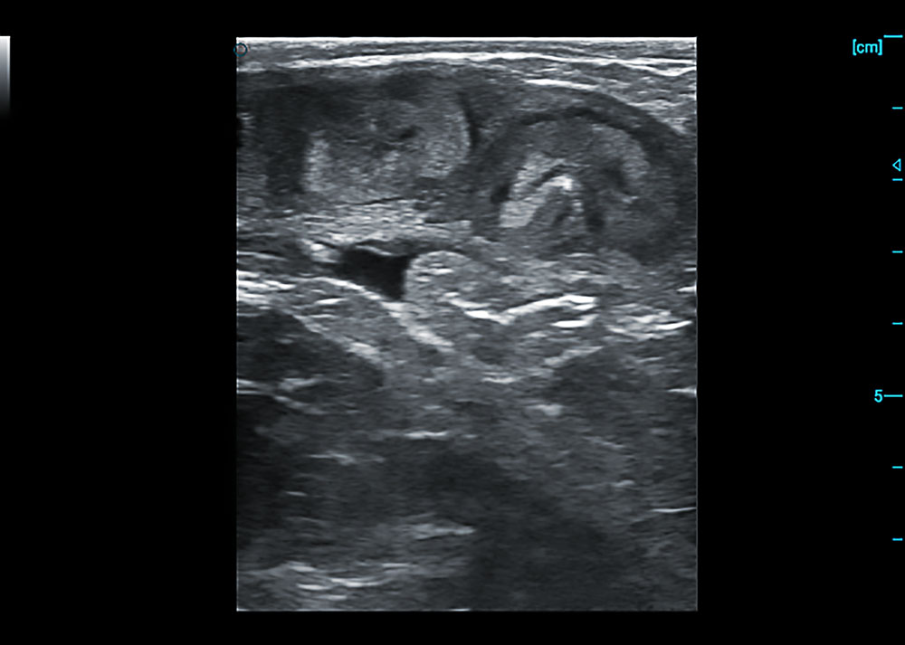
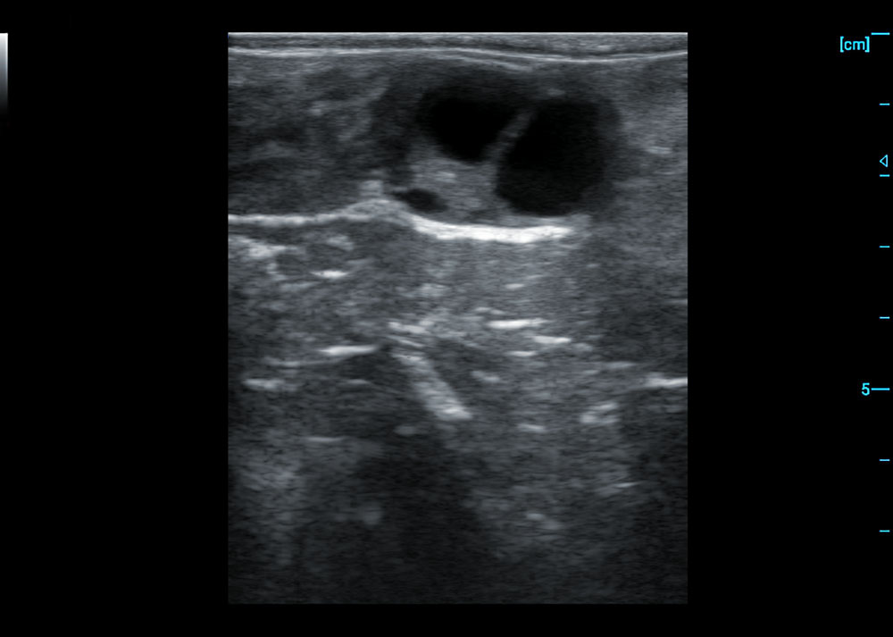
Non-Pregnant Uterus Imaging
• Detailed visualization of uterine anatomy including horn shape and lumen.
• Clearly discern endometrial layers for reproductive status evaluation.
• Effective tissue penetration for reliable imaging in cattle.
• Stable performance for consistent results across examinations.
-
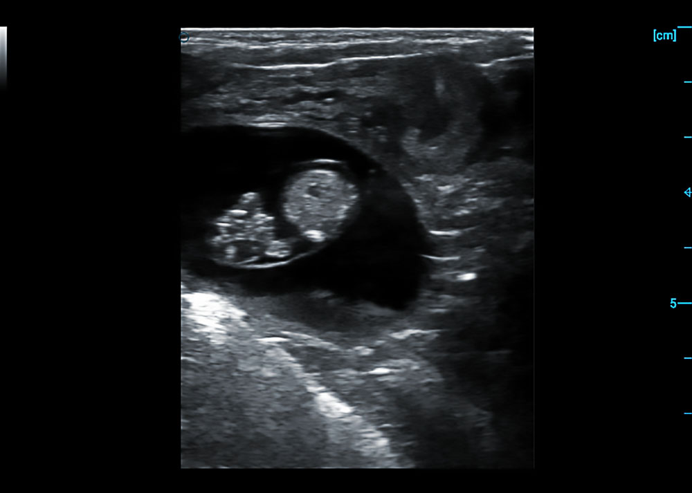
Early Pregnancy Confirmation
• Clear visualization of fetal heartbeat at 50 days gestation.
• Accurate measurement of crown-rump length for gestational age verification.
• Defined structural identification of early fetal development landmarks.
• Stable imaging performance in typical farm examination conditions.
-
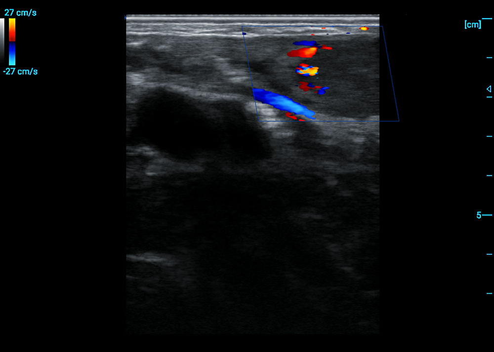
Ovarian Follicle Monitoring
• Multiple anechoic follicular structures clearly visualized with defined margins.
• Accurate measurement of dominant follicle diameter reaching 15mm.
• Distinct vascularization pattern detected via color Doppler assessment.
• Maintained image stability during transrectal probe manipulation.
-
Rectal Twin Pregnancy Scanning
• Simultaneous visualization of two distinct fetal heartbeats with cardiac activity.
• Precise biometric measurements confirming concordant growth between twins.
• Clear demarcation of separate chorioallantois membranes for each fetus.
• Color Doppler confirming adequate vascular supply to both gestational units.
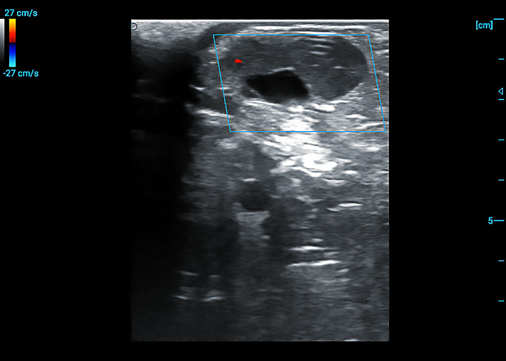
-
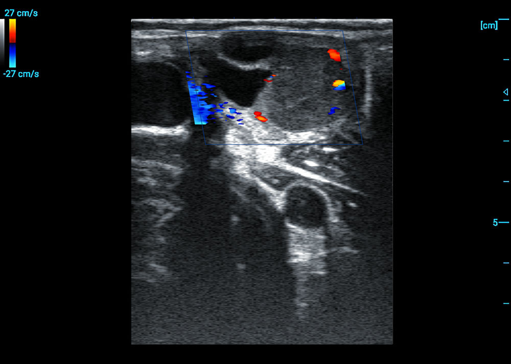
Corpus Luteum Visualization
• Well-defined hypoechoic structure with characteristic "stellate" central echo pattern.
• Active vascularization demonstrated by color Doppler with 27cm/s peak systolic flow.
• Precise measurement of luteal tissue diameter and vascular perfusion area.
• Stable hemodynamic imaging during transrectal examination procedure.

Demo Images
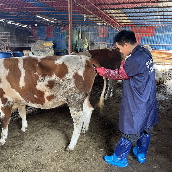
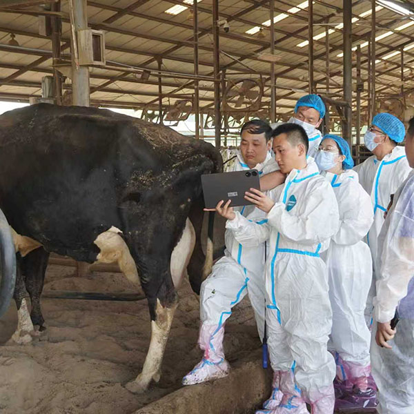
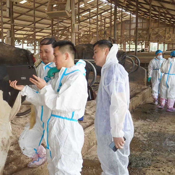
Operation Site