If you've ever watched a veterinarian move a probe over the side of a pregnant mare or listened to the gentle whooshing of blood flow from a cow’s uterine artery on a screen, you’ve already witnessed two different types of ultrasound technology in action. While both Doppler and B mode ultrasound rely on the same basic principle—using sound waves to create images—their purpose, output, and interpretation can be dramatically different.
")
Most people outside the veterinary or human medical field tend to lump all ultrasound into one category. But ask any experienced vet scanning a sheep’s ovary or tracking a foal’s heartbeat, and they’ll tell you: knowing the difference between Doppler and B mode ultrasound isn’t just academic—it shapes how we diagnose, monitor, and treat animals across a wide range of conditions.
Let’s break it down, practically and clearly, as if we were chatting over coffee at the clinic.
What is B Mode Ultrasound?
B mode (short for “Brightness mode”) ultrasound is the most commonly used imaging method in veterinary and human medicine. It creates a 2D grayscale image, allowing veterinarians to visually examine internal structures—muscles, organs, tendons, and even fetuses—in real time.
When you hear people talk about “ultrasound scans,” they’re usually referring to B mode. The machine sends out sound waves that bounce off tissues, and the echo patterns are translated into images where denser structures like bones show up white, and softer tissues appear darker.
What do vets use B mode for?
Checking pregnancy status in cows, mares, pigs, goats, and sheep
Examining ovaries, uterus, testicles, or kidneys
Evaluating muscle development and backfat in livestock
Detecting cysts, abscesses, or tumors
It’s particularly valuable because it’s non-invasive, quick, and repeatable. You can check a sow today, come back next week, and compare her reproductive status without any harm.
What does the image look like?
Imagine a black-and-white photo taken inside the body—where you can see different layers, fluid pockets, and organ boundaries. That’s B mode in action.
What is Doppler Ultrasound?
Doppler ultrasound goes a step beyond just structure. It focuses on movement, particularly the flow of blood. Named after the Doppler effect (the same phenomenon that makes a siren sound different as it moves past you), this technique measures changes in frequency caused by movement—like blood flowing through a vessel.
So instead of a static image, Doppler shows whether something is flowing, how fast it’s flowing, and in what direction.
There are three main types used in vet practice:
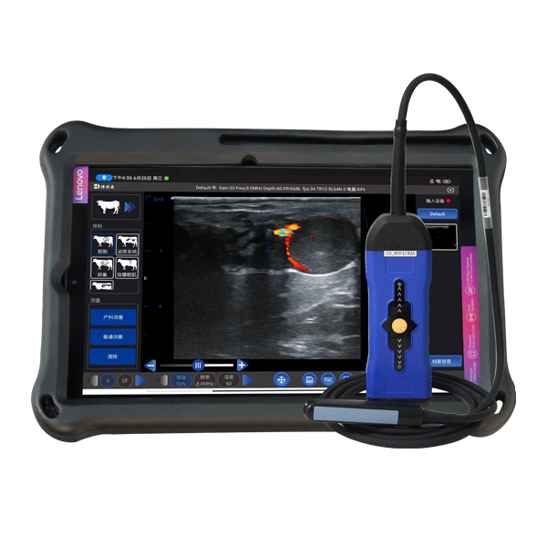
Color Doppler – Shows blood flow as color (usually red and blue) over a B mode image.
Power Doppler – More sensitive to low flow, useful in small vessels or reproductive organs.
Spectral Doppler – Graphs the speed of blood flow over time.
What do vets use Doppler for?
Monitoring fetal heart rates and umbilical blood flow
Detecting uterine artery health in cows or mares
Evaluating testicular blood flow in breeding bulls or boars
Identifying blood flow in tumors (is it benign or malignant?)
Checking circulation in injured limbs or organs
What does the image look like?
Overlay colors flowing through vessels, often on top of a B mode image—almost like a weather radar showing moving clouds.
Practical Example: Reproductive Exams in Livestock
Imagine you're checking the fertility of a breeding bull. A B mode scan shows the size, shape, and structure of his testicles. You can check for abnormalities like hypoplasia, mineralization, or trauma. But with Doppler, you can see the blood supply, which is directly related to semen quality.
Similarly, in a pregnant ewe, B mode gives a clear view of the fetus: its spine, heartbeat, even limb movement. But Doppler lets you hear and measure the blood flow through the umbilical cord, ensuring the fetus is getting enough oxygen and nutrients. If that blood flow is restricted, it’s a sign of trouble.
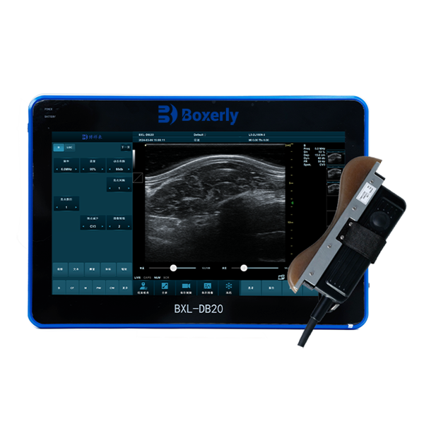
That’s why many modern portable ultrasound systems for livestock, like the BXL-V50, are equipped with both modes—they let you assess both structure and function in a single scan.
")
Which One Should Be Used—and When?
The choice between Doppler and B mode isn’t “either-or”—they complement each other.
B mode is your first-line tool. It’s ideal for routine exams, structural evaluations, and initial diagnosis.
Doppler becomes essential when function or blood flow is critical to evaluate, especially in advanced reproductive monitoring or circulatory assessments.
Vets working in large animal reproduction often use B mode to identify follicles, corpora lutea, or pregnancy status, then switch to Doppler to measure luteal blood flow—a key indicator of whether ovulation and progesterone production are healthy.
Interpreting the Results: What’s the Learning Curve?
Here’s where things get interesting. B mode is relatively beginner-friendly. With basic training, most vets or trained techs can start identifying reproductive structures or pregnancy in livestock.
Doppler takes a bit more finesse. Because it deals with flow direction, speed, and waveform patterns, interpretation can be more subjective and takes practice.
Also, Doppler needs better probe handling and machine settings—angle, pressure, and positioning all affect the result. That’s why clinics introducing Doppler features usually invest in additional training.
Why Does This Matter on a Farm?
It might sound like high-tech theory, but here’s how the difference between Doppler and B mode plays out in the field:
A sow with sluggish umbilical blood flow might lose her litter. Doppler catches that.
A cow with a large follicle but no blood flow? It might not ovulate—breeding should be delayed.
A bull with good testicle size but poor perfusion? He's likely subfertile, despite appearances.
A foal with abnormal blood flow from the placenta could be at risk. Early intervention saves lives.
On a commercial farm, time is money. Being able to distinguish between a healthy pregnancy and a compromised one helps adjust management strategies early—feed, medicine, isolation, or re-breeding decisions are made with actual data, not guesswork.
The Equipment Side
Today’s portable Veterinary ultrasound systems are much more advanced than a decade ago. Units like the BXL-V50 integrate both B mode and Doppler in a compact, battery-powered device that’s designed for field use. They’re durable, weather-resistant, and work with a wide range of probes—linear, convex, transrectal—so you can switch between a mare, a cow, and a boar without changing machines.
The rise in wireless connectivity and real-time image sharing also makes these tools accessible to more farms, not just large breeding operations. You can scan in the barn and consult with a specialist via phone right away.
Key Takeaways
Let’s wrap up the difference in plain terms:
| Feature | B Mode Ultrasound | Doppler Ultrasound |
|---|---|---|
| Focus | Structure (shape, size, anatomy) | Movement (mainly blood flow) |
| Image type | Grayscale 2D | Color overlay or waveform |
| Common Uses | Pregnancy checks, organ scans | Blood flow, fetal health, vascular status |
| Learning curve | Moderate | Higher |
| Tools used | Standard probe | Doppler-capable probe and settings |
| Field applications | Reproduction, growth, injuries | Fertility, fetal oxygenation, circulatory health |
If you work with livestock, combining both B mode and Doppler gives you a complete picture—you don’t just see what’s there, you understand how well it’s working.
And in animal health, that difference can mean a successful pregnancy, a stronger breeding program, or simply less guessing and more clarity in your daily work.