Pyometra in female dogs is often more obvious 4-10 weeks after the late estrus. The vulva of the sick dog is thickened and swollen, and the dog continues to estrus and bleed. The dog is depressed, has a decreased appetite, and has less activity. As the disease progresses, the abdomen is sensitive to palpation, and the body temperature rises. The appetite gradually disappears, the desire to drink gradually increases, and the dog gradually loses weight. Some sick dogs vomit. The disease progresses rapidly with a swollen abdomen, and can eventually lead to shock and death. Veterinary B-ultrasound should be used frequently to check the condition of the uterus in the late estrus.
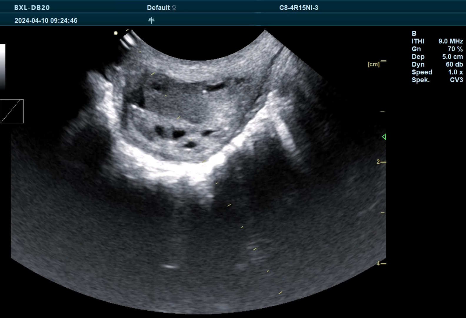
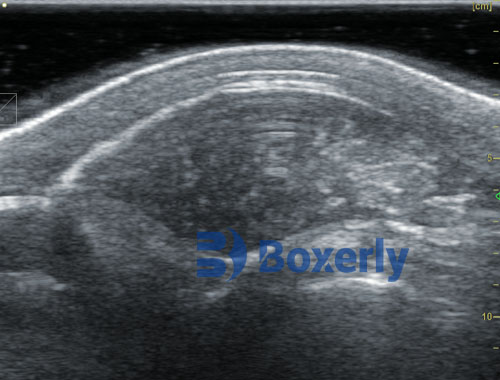
Closed pyometra, the cervix is completely closed and blocked, there is no purulent secretion from the vulva, the abdominal circumference is large, the breathing and heartbeat are accelerated, and in severe cases, breathing is difficult, the abdominal skin is tense, the subcutaneous veins in the abdomen are dilated, and the dog likes to lie on the ground. When using VETERINARY B-ULTRASOUND for observation, closed pyometra is a large low-echo area on the veterinary B-ultrasound.

Open uterine pyometra, veterinary B-ultrasound can be observed that the cervix is not completely closed, and a small amount of purulent secretions flow out from the vulva from time to time, which are cheese-like, creamy yellow, gray or reddish brown, odorless or with a strong fishy smell. The dog's vulva is red and swollen, the vaginal mucosa is flushed, and the abdominal circumference is slightly enlarged. Its status can be easily observed on veterinary B-ultrasound.
Veterinary X-ray examination, hematological examination and ultrasound examination can determine whether the uterus has pus. Veterinary X-ray examination can show an enlarged uterus (closed or open); veterinary B-ultrasound examination can show turbid fluid in the uterus and thinning of the uterine wall (excluding ascites, pregnancy, tumor); blood test shows slight or significant increase in white blood cells, increase in granulocytes, anemia, and increase or decrease in platelets.
link: https://www.bxlimage.com/nw/211.html
tags: veterinary B-ultrasound Veterinary Ultrasound


