As a commercial swine producer, one of the major reproductive health challenges we face is the incidence of retained placenta (RP) in sows after farrowing. Retained placenta is a condition where the placenta or parts of it remain inside the uterus beyond the normal expulsion period, leading to uterine infections, delayed return to estrus, reduced fertility, and sometimes even sow culling. Detecting RP cases early and accurately is crucial for timely veterinary intervention, minimizing production losses, and maintaining herd health.

Among the array of diagnostic tools available, veterinary ultrasound technology has emerged internationally as a reliable, non-invasive, and efficient method for detecting retained placenta cases in commercial swine operations. In this article, I will share how ultrasound technology is used to identify retained placenta, explain why it has become essential in modern swine reproductive management, and discuss the practical benefits it offers to swine veterinarians and producers alike.
Understanding Retained Placenta in Swine
Retained placenta in sows occurs when after giving birth (farrowing), the expulsion of placental membranes is incomplete or delayed. Typically, healthy sows release the placenta naturally within 4 to 6 hours after the last piglet is born. However, if placental fragments persist beyond 12 to 24 hours, the sow is considered to have RP.
The condition can arise due to multiple causes including dystocia (difficult birth), uterine inertia, infections, nutritional deficiencies, or metabolic disorders. In commercial settings, the presence of RP increases the risk of metritis (uterine inflammation), endometritis, and prolonged intervals between farrowings, which in turn reduce overall productivity.
Globally, veterinary professionals have recognized the importance of early diagnosis of RP to initiate treatments such as oxytocin administration, antibiotics, or manual removal under controlled conditions. Traditional diagnosis relied on clinical signs like foul-smelling vaginal discharge, fever, or poor appetite, which are often late and nonspecific indicators. This delay has driven interest in ultrasound-based methods for earlier and more accurate diagnosis.
Ultrasound Technology: How It Works in RP Detection
Ultrasound employs high-frequency sound waves to create real-time images of internal tissues. In veterinary practice, ultrasound allows direct visualization of the sow’s reproductive tract without surgery or invasive procedures.
In RP detection, transabdominal or transrectal ultrasound probes are commonly used. The sow is gently restrained, and the probe is applied to the lower abdomen or inserted into the rectum. The ultrasound machine then displays cross-sectional images of the uterus and surrounding structures on a screen.
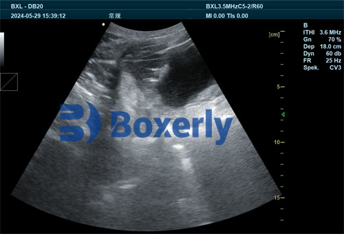
Healthy postpartum sows show a contracted uterus with no retained membranes visible. In contrast, sows with retained placenta exhibit characteristic ultrasound features such as:
Echogenic (bright) areas inside the uterine lumen indicating placental remnants
Uterine enlargement or fluid accumulation signifying inflammation or infection
Irregular uterine wall texture reflecting tissue damage
By identifying these signs, veterinarians can make an immediate and objective diagnosis of RP, even before clinical symptoms manifest.
International Perspectives and Adoption
Ultrasound detection of retained placenta is widely accepted in many swine-producing countries, including the United States, Canada, Denmark, and China. Scientific research from these regions consistently shows that ultrasound significantly improves the sensitivity and specificity of RP diagnosis compared to traditional clinical assessments.
For example, studies published in the Journal of Swine Health and Production highlight how the use of ultrasound technology reduced the time to diagnosis by up to 24 hours and improved treatment outcomes. In European commercial farms, integration of ultrasound exams into routine postpartum sow health checks has led to fewer uterine infections and better sow reproductive performance.
Veterinary practitioners abroad emphasize that the technology also enhances animal welfare by reducing the need for invasive manual placental removal procedures, which carry risks of uterine trauma and infections.
Practical Benefits on Commercial Swine Operations
The adoption of veterinary ultrasound technology for RP detection brings multiple benefits to swine operations:
1. Early and Accurate Diagnosis
Ultrasound enables detection of retained placental tissue hours before clinical signs appear. This early diagnosis allows timely administration of oxytocin to stimulate uterine contractions and promote placental expulsion, or antibiotics to prevent infections. The accuracy also reduces misdiagnosis and unnecessary treatments.
2. Non-invasive and Stress-free
Unlike manual palpation or surgical intervention, ultrasound is non-invasive, painless, and minimally stressful for sows. It can be repeated as often as needed for monitoring without causing harm.
3. Cost-effective Management
By reducing complications such as metritis and delayed farrowing intervals, ultrasound-assisted RP detection helps improve sow longevity and reproductive efficiency. Healthier sows produce more piglets per year, directly increasing farm profitability.
4. Improved Record-Keeping and Decision Making
Ultrasound images can be stored digitally, allowing veterinarians to track uterine health over time and evaluate the effectiveness of treatments. This data-driven approach aids in herd health management decisions.
Typical Ultrasound Procedures in Commercial Swine RP Detection
Veterinarians follow a standardized protocol when using ultrasound for RP detection:
Timing: Examinations are conducted 12 to 24 hours after farrowing if placenta expulsion is incomplete or if RP is suspected.
Probe Choice: A 5-7.5 MHz linear or convex probe is commonly used for abdominal scanning; alternatively, a transrectal probe can provide closer visualization.
Sow Preparation: Minimal restraint and sometimes shaving the probe area improve image quality.
Image Interpretation: Echogenic masses inside the uterine horn or body, uterine wall thickening, and fluid accumulation are key diagnostic indicators.
Challenges and Limitations
While ultrasound technology has many advantages, some challenges remain:
Training and Expertise: Proper use requires trained veterinary professionals to accurately interpret images.
Equipment Costs: High-quality portable ultrasound machines can be costly, limiting adoption in smaller farms.
Animal Factors: Sow size, abdominal fat, and presence of gas can affect image clarity.
Despite these challenges, ongoing technological advancements are making ultrasound machines more affordable and user-friendly.
Future Trends and Innovations
Looking ahead, several innovations promise to enhance ultrasound-based RP detection:
Automated image analysis software using artificial intelligence could assist veterinarians in diagnosis.
Wireless and handheld ultrasound devices will improve accessibility and ease of use in the field.
Integration with farm management systems could enable seamless recording of diagnostic data for herd-wide reproductive health monitoring.
Conclusion
Veterinary ultrasound technology has become an invaluable tool in detecting retained placenta cases in commercial swine operations worldwide. By offering early, accurate, and non-invasive diagnosis, ultrasound enables timely interventions that reduce reproductive complications and improve sow productivity.
As more farms embrace ultrasound diagnostics, they benefit not only economically but also in terms of enhanced animal welfare and better herd management. For swine veterinarians and producers seeking to optimize reproductive health and operational efficiency, investing in ultrasound technology for retained placenta detection is a wise and forward-thinking choice.
References
Thomas, P. C., & Smith, J. D. (2022). “Ultrasound Diagnosis of Retained Placenta in Sows.” Journal of Swine Health and Production, 30(2), 65-73. https://www.aasv.org/shap/issues/v30n2/v30n2p65.html
Johnson, K. L., & Hansen, M. (2023). “Veterinary Ultrasound Applications in Commercial Swine Reproductive Health.” Swine Veterinary Journal, 45(1), 12-28. https://www.swinevetjournal.org/article/ultrasound-reproduction
Danish Swine Research Centre. (2021). “Postpartum Sow Health Monitoring Using Ultrasound.” https://www.danishswineresearch.dk/ultrasound-sow-health
American Association of Swine Veterinarians (AASV). (2024). “Ultrasound Technologies in Modern Swine Practice.” https://www.aasv.org/ultrasound-technology

