Understanding the internal health and reproductive status of animals has always been a cornerstone of modern veterinary medicine. Among the many diagnostic tools available today, veterinary ultrasonography stands out as a non-invasive, safe, and dynamic method. As veterinary care evolves, this technology has increasingly become an integral part of livestock management, equine medicine, companion animal care, and reproductive diagnostics. However, despite its many advantages, there are still challenges in application that need to be addressed. In this article, we explore both the limitations and future prospects of animal ultrasonography, incorporating insights from international veterinary practice to highlight its global significance.
")
Practical Issues in Animal Ultrasonography
1. Interpretation Requires Expertise
Although ultrasonography can vividly display internal anatomical structures such as the ovaries, uterus, liver, kidneys, and even fetal organs, its diagnostic accuracy is highly dependent on the operator’s skill. Unlike laboratory tests that yield numerical results, ultrasound images are real-time, greyscale visuals that must be interpreted by the veterinarian. This means extensive training is necessary to differentiate between normal and pathological findings.
For example, ovarian cysts and normal follicles may appear similar in size and shape under ultrasound. Misinterpretation can lead to inappropriate treatments or missed diagnoses. Thus, foreign veterinarians emphasize the importance of continuous training, often through certification programs such as those offered by the American College of Theriogenologists or the European Veterinary Diagnostic Imaging societies.
2. B-Mode Imaging Has Limits
The most common ultrasound mode used in animals is B-mode (brightness mode) imaging. While excellent for detecting structural changes and general shapes, it cannot provide detailed information about blood flow, tissue elasticity, or metabolic activity. In complex cases—such as differentiating between a malignant and benign uterine mass—additional tools such as Doppler ultrasonography, elastography, or CT/MRI may be required.
3. Ultrasonography Is Not a Standalone Diagnostic Tool
Veterinary experts across Europe, North America, and Australia emphasize that ultrasonography should be combined with other diagnostic methods. Clinical history, physical examination, blood tests, hormone assays, and even biopsy may be needed for a comprehensive diagnosis. For instance, in dairy cattle, ultrasonography is often paired with blood progesterone testing to accurately diagnose corpus luteum cysts. A Chinese study found ultrasound had a 92.6% accuracy rate, while blood hormone tests ranged from 70–80%—supporting the value of combining methods for higher reliability.
4. Equipment and Maintenance Costs
While ultrasonography units are becoming more affordable, high-resolution portable veterinary machines still represent a significant investment for many farms or small clinics. Additionally, regular maintenance and technological updates are needed to ensure optimal performance. This cost barrier is particularly prominent in developing countries, though international aid and veterinary associations are working to increase access through training and subsidized equipment programs.
")
Global Progress in Veterinary Ultrasonography
Despite these challenges, the development of animal ultrasonography has been remarkable. In the last two decades, improvements in image quality, probe flexibility, and digital data storage have enhanced both diagnostic accuracy and clinical workflow.
1. Safer and Repeatable Diagnostics
Unlike radiographic imaging, ultrasonography poses no radiation risk, making it ideal for repeated examinations—especially during pregnancy or post-surgical recovery. Veterinarians in the UK and Canada frequently use ultrasonography to monitor uterine involution in postpartum cows, track fetal growth, or examine equine tendon injuries over time.
2. Advancing Reproductive Management
Veterinary ultrasonography has revolutionized reproductive monitoring. In breeding farms worldwide—especially in the U.S., Australia, and Germany—this tool is widely used to detect early pregnancy, monitor embryonic viability, and guide artificial insemination or embryo transfer programs. For instance, in swine and sheep breeding, accurate timing of insemination using ultrasound-guided follicular measurements has significantly improved conception rates.
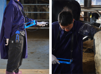
3. Portable and Field-Friendly Devices
Modern portable scanners, such as wireless ultrasound probes, have made it easier for veterinarians to conduct examinations on-farm, in wildlife reserves, or in remote areas. In the U.S. and New Zealand, portable ultrasonography is widely used by large-animal veterinarians and wildlife conservationists to monitor health conditions in cattle, deer, and even marine mammals.
")
Promising Directions in Ultrasonography
The future of veterinary ultrasonography is not just about better images—it’s about intelligent diagnostics, integration with AI, and global accessibility. Here’s where the field is heading:
1. AI-Enhanced Image Analysis
Several international research teams, including those in Germany and South Korea, are developing AI algorithms to assist in real-time image analysis. These systems help flag abnormalities, calculate organ dimensions, and even predict fetal age. This could greatly reduce operator dependency and make accurate diagnostics available to less experienced users.
2. Integration with Herd Management Systems
Some advanced livestock farms in the U.S. and Denmark are integrating ultrasonographic data into herd management software. For example, cow ovarian status, pregnancy stage, and uterine health can be uploaded and analyzed alongside milk yield and feed intake data, allowing for precision reproductive and nutritional planning.
3. Educational Expansion
Veterinary schools around the world are expanding their ultrasonography training modules. In Europe, the European School of Advanced Veterinary Studies offers specialized certificates, while in the U.S., universities such as Cornell and UC Davis are integrating handheld ultrasound training from the first year. This ensures that future veterinarians will be confident in using this tool from early in their careers.
4. Cross-Species Applications
Veterinary ultrasonography is no longer limited to food-producing animals. Companion animals (cats, dogs), exotic pets (rabbits, reptiles), and even zoo animals are now being scanned regularly. For instance, ultrasonography has become standard in canine and feline pregnancy monitoring, as well as in diagnosing gastrointestinal obstructions, heart disease, or renal disorders.
")
Conclusion
Veterinary ultrasonography has transformed the diagnostic landscape in animal health. Though it presents challenges—such as requiring skilled interpretation and being limited in certain pathologies—its advantages in safety, real-time imaging, and diagnostic value make it indispensable. As technology advances, especially with AI and portable innovations, ultrasonography is poised to play an even more central role in global veterinary medicine.
Incorporating insights from both developed and developing countries, we see a shared vision: to make veterinary ultrasonography more accessible, more accurate, and more integral to daily animal care. For livestock producers, pet owners, and veterinarians alike, this technology represents not only a window into the animal’s body but also a tool for better decision-making, improved animal welfare, and more sustainable veterinary practice.