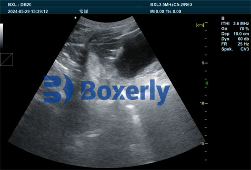
In modern pig production, the efficiency and profitability of a breeding herd depend heavily on sow reproductive performance. Two critical challenges that significantly impact farm economics are the duration of non-productive days (NPDs)—commonly referred to as “empty days” between weaning and successful conception—and improper backfat thickness in sows. Fortunately, veterinary ultrasonography has emerged as an essential diagnostic and management tool to address both.

In this article, we will explore how ultrasonography helps reduce NPDs and optimize sow backfat, drawing on practices widely recognized by veterinarians and producers in Europe, North America, and Asia.
Understanding Non-Productive Days (NPDs)
Non-productive days represent the time a sow is neither pregnant nor lactating. These periods are costly because the animal continues to consume resources without generating income through piglet production. A high NPD rate is often indicative of poor heat detection, delayed breeding, failed insemination, or underlying reproductive disorders.
Veterinary ultrasonography helps detect these issues early. By using a B-mode ultrasound scanner, producers can visualize ovarian follicles, corpora lutea, uterine fluid, or early pregnancy signs. This information allows for timely breeding decisions and correction of reproductive pathologies.
Gestation Length in Sows and the Importance of Timing
The average gestation length in sows is 114 days, although it may range from 111 to 117 days. Breeds like Yorkshire and Landrace may vary slightly in gestation duration, but the difference is generally within a narrow margin (e.g., 114.34 days for Yorkshire and 114.38 days for Landrace).
These small differences underline an important point: the farm’s reproductive management practices—not just genetics—are critical to performance. By managing NPDs through early pregnancy diagnosis and reproductive evaluation using ultrasound, farms can maintain regular farrowing intervals and improve overall reproductive efficiency.
Using Ultrasonography to Detect Early Pregnancy
One of the most widespread uses of ultrasound in swine production is early pregnancy diagnosis. Typically, a transabdominal B-mode scanner is used between days 21 and 30 post-breeding. A trained operator can identify the presence of fluid-filled embryonic vesicles in the uterus as early as day 24.
Confirming pregnancy early has several benefits:
Non-pregnant sows can be identified quickly and re-bred without wasting time.
Management decisions can be adjusted to avoid prolonged empty periods.
Resources such as housing and feed can be better allocated based on reproductive status.
In many European and North American pig farms, integrating routine ultrasound checks into breeding protocols has helped reduce NPDs to below 12 days per sow per year—considerably lower than global averages.

Managing Backfat Thickness for Optimal Reproductive Success
Another essential application of ultrasonography is the measurement of sow backfat thickness. Backfat, measured at the P2 position (approximately 6.5 cm from the midline at the last rib), is a reliable indicator of a sow’s energy reserves. Both excessive and insufficient backfat can lead to reproductive inefficiencies.
Sows with too little backfat (<14 mm) often experience:
Delayed return to estrus after weaning
Lower conception rates
Reduced litter sizes
On the other hand, sows with excessive backfat (>20 mm) may suffer from:
Difficult farrowing (dystocia)
Higher risk of stillbirths
Decreased feed intake during lactation
Veterinary ultrasonography enables precise, repeatable measurement of backfat. With real-time data, producers can fine-tune feeding strategies during gestation and lactation to ensure that sows maintain an optimal body condition.
Post-Weaning Monitoring and Timely Breeding
One of the most critical windows in sow reproduction is the interval between weaning and successful breeding. Ideally, sows should come into estrus and be bred within 5–7 days after weaning. If this window is missed, it can lead to extended NPDs and higher production costs.
Ultrasound scanning of reproductive organs shortly after weaning can reveal:
Whether the ovaries are developing follicles
The presence of uterine fluid or endometritis
General uterine involution status
If a sow fails to return to estrus, ultrasound can assist in differentiating between ovarian inactivity, silent estrus, or pathological conditions. Interventions such as hormone therapy or nutritional support can then be promptly administered.
Balancing Reproductive Recovery and Efficiency
Some researchers and practitioners have observed that a slightly extended weaning-to-estrus interval allows reproductive organs more time to recover, particularly in sows that experienced stress or poor body condition during lactation. However, this must be carefully balanced against the need to minimize non-productive days.
Ultrasound plays a central role here by tracking uterine and ovarian recovery. Rather than relying on external signs alone, farmers can use objective ultrasonographic data to determine the optimal breeding time for each sow.
Integrating Ultrasound into Routine Reproductive Management
In well-managed farms, veterinary ultrasonography is used not only for pregnancy detection but also for routine monitoring of reproductive health. A few examples of ultrasound-assisted decisions include:
Identifying sows with cystic ovaries or prolonged luteal phases
Verifying complete uterine involution after farrowing
Selecting gilts with well-developed ovaries for breeding
Monitoring backfat changes during gestation
These insights reduce guesswork, shorten decision cycles, and allow interventions to be timely and precise.
Economic Benefits of Reducing NPDs and Backfat Imbalances
Reducing NPDs and optimizing backfat through ultrasound translates directly into financial gain:
A reduction of just 1 NPD per sow can increase farm income by $5–$10 per sow per year, depending on the market.
Maintaining optimal backfat improves litter performance, reduces stillbirths, and shortens the interval to rebreeding.
Efficient breeding reduces the number of inseminations per pregnancy, lowering semen and labor costs.
Real-World Adoption of Ultrasonography
In countries like Denmark, the Netherlands, and Canada—known for their high-efficiency pig farming systems—veterinary ultrasonography is considered standard practice. Farms have dedicated technicians or veterinary teams who routinely scan sows during critical points in the reproductive cycle.
In the United States, portable and wireless ultrasound devices are increasingly popular. These allow real-time scanning in the pen, reducing stress on animals and improving workflow efficiency.
In Asia, where pig density is high and space is limited, ultrasound helps producers reduce non-productive time and manage body condition to ensure better turnover of sows.
Future Outlook: Automation and Precision Reproduction
As precision livestock farming evolves, ultrasonography is being integrated with AI and farm management software. Smart ultrasound probes that can transmit images to cloud-based databases are becoming more common. These innovations promise more automated tracking of reproductive cycles, allowing even faster decisions on breeding or culling.
Ultrasound data can also be linked with other sensor data (e.g., feeding behavior, weight, and temperature) to create a full profile of sow health and productivity. This holistic approach will become increasingly important as farms aim to produce more with fewer resources.
Conclusion
Veterinary ultrasonography is no longer a luxury or an occasional diagnostic tool—it is a cornerstone of reproductive management in modern pig production. By reducing non-productive days and maintaining optimal backfat thickness, farms can improve sow performance, reduce costs, and enhance overall herd productivity.
As more producers around the world adopt ultrasound technology and integrate it into their daily routines, the result will be not only more efficient operations but also better animal welfare and sustainability outcomes. For any pig farm aiming to remain competitive, investing in ultrasonographic expertise and equipment is a step in the right direction.


