In modern dairy farming, reproductive efficiency is a cornerstone of profitability. Early detection of ovarian activity and abnormalities in dairy cows plays a critical role in improving calving intervals, optimizing breeding strategies, and enhancing herd fertility. Veterinary ultrasonography—specifically B-mode ultrasound imaging—has become a gold standard tool for evaluating the female reproductive tract in cattle. Among the many techniques available, two primary methods are widely adopted when using Veterinary ultrasound to examine dairy cow ovaries: the transvaginal method and the transrectal method.
")
Each approach provides unique advantages, and both are extensively used in practice across many parts of the world, especially in North America, Europe, and Australia. In this article, we will explore these two approaches in detail, highlight their applications in different reproductive stages, and discuss how veterinarians and livestock producers utilize this technology to manage herd fertility effectively.
Understanding Veterinary Ultrasound in Dairy Cow Reproduction
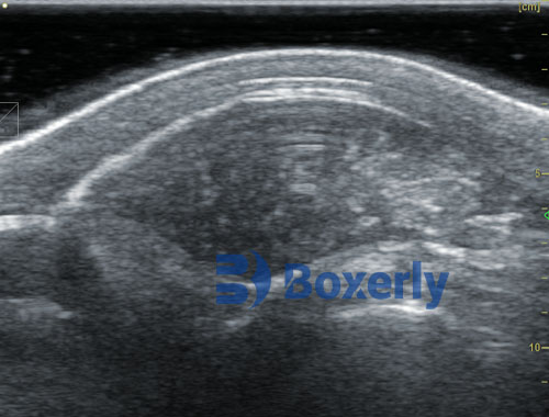
Veterinary ultrasound, particularly B-mode real-time imaging, allows for non-invasive, accurate, and immediate observation of the internal reproductive structures of cattle. This includes the uterus, ovaries, follicles, corpus luteum (CL), and early pregnancy status. Compared to traditional rectal palpation, ultrasound imaging offers significantly better sensitivity and specificity, and it enables the detection of small ovarian structures that might otherwise go unnoticed.
1. The Transvaginal Ultrasonographic Method
The transvaginal approach involves introducing a specially designed ultrasound probe into the vaginal canal. This technique is primarily utilized for detailed observation of the lower reproductive tract, including the cervix, uterine body, and ovaries.
Procedure:
The veterinary ultrasound probe is slowly inserted into the cow’s vaginal canal and carefully advanced toward the vaginal fornix. The probe’s position is then adjusted vertically and laterally to achieve a clear image of the ovarian structures.
Advantages:
Offers close proximity to the ovaries and uterine horns
Particularly useful for diagnosing ovarian cysts or abnormalities close to the vaginal wall
Ideal for fixed cattle in restraint systems, where precision is possible
Often used in reproductive research or embryo collection settings
Limitations:
Requires a high level of operator skill and hygiene to avoid contamination
Less comfortable for the animal than the transrectal approach
May not be suitable for routine use on large farms due to time constraints
Application in Practice:
While less commonly used in day-to-day reproductive management, the transvaginal method is favored in embryo transfer programs, controlled breeding experiments, and ovarian puncture procedures for oocyte aspiration. In some research centers in the U.S. and Europe, transvaginal ultrasound-guided follicular aspiration is a routine part of in vitro embryo production protocols.
")
2. The Transrectal Ultrasonographic Method
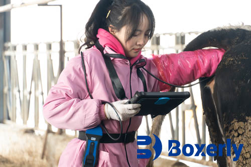
By far the most common method used worldwide, the transrectal approach involves inserting a short veterinary ultrasound probe into the rectum. The probe is positioned against the rectal wall, which lies adjacent to the uterus and ovaries, allowing the operator to scan these structures through the intestinal wall.
Procedure:
The rectum is gently cleared of fecal material, and the ultrasound probe—usually mounted on a stiff handle—is guided manually into the rectum. The operator then manipulates the probe to obtain images of the uterus and both ovaries, examining for follicles, CL, or signs of pregnancy.
Advantages:
Widely used due to its practicality and minimal discomfort for the cow
Can be performed quickly and efficiently in a herd setting
Allows detailed observation of follicles, CL development, and pregnancy status
Suitable for early detection of ovarian disorders and silent estrus
Limitations:
May be slightly less precise than the transvaginal method for very small structures
Requires proper training to avoid misdiagnosis or injury
Challenging in very young heifers or postpartum cows with inflamed reproductive tracts
Global Usage and Adoption:
In countries such as the United States, Canada, Germany, and Australia, the transrectal ultrasound has become standard practice in reproductive herd health programs. Many large-scale dairy operations schedule regular ultrasound checks to monitor post-calving ovarian activity, confirm pregnancy, and assess reasons for infertility.
Clinical Cases: Silent Estrus and Short Luteal Phase Detection
Ultrasound imaging is especially valuable in identifying reproductive anomalies that are not externally visible. For instance:
1.Silent Estrus (Anestrus with Ovarian Activity):
In dairy cows after parturition, progesterone levels typically rise around 28 days postpartum, peaking around 34 days at about 3 ng/mL. During this time, the cow may not exhibit external signs of estrus, yet the ovary displays luteal activity (confirmed by a CL on ultrasound). Progesterone declines slightly until around 48 days, when an anovulatory follicle may be present. Ovulation may finally occur around 72 days postpartum, when progesterone rises again, peaking at 3.6 ng/mL and visible CL returns.
2.Short Luteal Phase:
Some cows exhibit a shortened luteal cycle between 62–72 days postpartum. Ultrasound examination during this window often reveals a small CL of short duration. This reproductive pattern is critical to identify, as such cows may return to estrus sooner than expected, leading to mistimed inseminations.
Veterinary ultrasound allows these reproductive profiles to be diagnosed in real time and with high accuracy, enabling interventions such as timed artificial insemination (TAI), hormone synchronization, or supportive nutritional and therapeutic treatments.
Why Veterinary Ultrasound Is Essential in Reproductive Management
Across advanced dairy industries, veterinary ultrasound is not merely a diagnostic tool—it is a decision-making instrument. Here’s why:
Non-invasive and repeatable: Repeated scans can monitor follicle growth, CL regression, or pregnancy development without harming the cow.
Economic return: Timely diagnosis of non-pregnancy or ovarian dysfunction allows for re-breeding strategies, reducing open days and improving calving intervals.
Enhancing reproductive technologies: Programs like embryo transfer and in vitro fertilization rely heavily on ultrasound to guide follicle aspiration and uterine evaluation.
Disease management: Ultrasound helps identify uterine infections, cystic ovaries, and other causes of reproductive inefficiency.
Future Outlook: Integration with AI and Data Systems
With technological advancements, veterinary ultrasound is now integrating with herd management software, allowing veterinarians to upload real-time imaging data into digital records. In countries like the Netherlands and New Zealand, AI-powered image recognition tools are being piloted to automatically detect ovarian phases and suggest breeding windows.
Moreover, portable wireless ultrasound probes that connect to smartphones or tablets are making this technology more accessible to smallholder farmers and rural veterinarians, broadening the global impact.
Conclusion
Veterinary ultrasound has revolutionized the reproductive management of dairy cows. The transvaginal and transrectal approaches both offer reliable access to ovarian structures and provide invaluable information for improving fertility outcomes. While transrectal ultrasound remains the most practical and widely used method, the transvaginal technique serves a critical role in advanced reproductive procedures.
By integrating these ultrasound techniques into routine herd health management, dairy producers can make informed decisions, optimize breeding efficiency, and enhance overall herd productivity. As the dairy industry continues to modernize, veterinary ultrasound will remain at the heart of reproductive success.