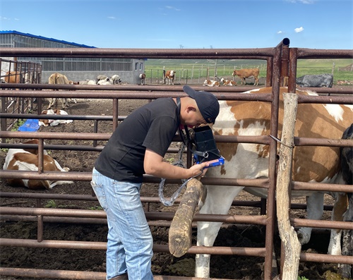
In today’s livestock veterinary medicine, ultrasound has become one of the most indispensable imaging tools—not only for reproductive assessments but increasingly for evaluating organ health, particularly the liver. Beyond just providing grayscale images, Veterinary ultrasound now offers advanced image texture analysis capabilities that can detect subtle changes in tissue architecture. This article explores how texture analysis of veterinary ultrasound images—especially in bovine liver—enhances disease detection, and how methods such as histogram analysis, gray-level co-occurrence matrices, and wavelet decomposition are transforming veterinary diagnostics.
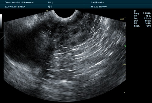
Understanding Ultrasound Texture in Veterinary Medicine
Ultrasound imaging generates grayscale pictures based on how tissues reflect high-frequency sound waves. When examining the liver or other internal organs of cattle, the distribution of echo intensity and its spatial variations result in what is known as "texture." In simpler terms, texture refers to the pattern or feel of an image area—it may appear coarse, fine, smooth, or irregular depending on the tissue's properties.
The texture of a bovine liver ultrasound is not random. It arises from the interaction of ultrasound pulses with different fibrous structures, fat content, and vascular architecture within the liver. For instance, a liver suffering from fatty infiltration (hepatic lipidosis) reflects ultrasound waves differently than a healthy liver, resulting in a visibly altered image texture. These variations can be quantified and analyzed through computational techniques, making ultrasound not just a visual tool but a measurable diagnostic platform.
Defining Texture in Ultrasound Imaging
In imaging science, texture is defined as the repetition of certain patterns or pixel arrangements, which can be either highly regular (e.g., a brick wall) or random but statistically consistent (e.g., sand or wood grain). In veterinary ultrasound, the liver’s echotexture might show subtle irregularities caused by pathological changes—yet these are often difficult to detect by eye alone. That’s where texture analysis comes into play.
Texture analysis uses image processing techniques to extract numerical features that describe the spatial organization of pixels. These features can then be used to classify tissues or detect abnormalities. In veterinary applications, especially cattle diagnostics, such analysis is essential for detecting liver dysfunctions that are not immediately visible to the naked eye during real-time scanning.
Four Primary Texture Analysis Methods
Veterinary researchers and practitioners commonly use four categories of texture analysis:
Statistical Methods
These involve calculating statistical measures from the distribution and relationships of pixel intensities. For veterinary ultrasound, this approach is particularly effective for irregular or visually ambiguous textures like fatty liver. Key statistical techniques include:
Gray-Level Histogram: Shows the frequency of each brightness level across the image. A liver with high fat infiltration often displays a shifted histogram due to increased brightness.
Gray-Level Co-occurrence Matrix (GLCM): Measures how often pairs of pixel values occur at specific distances and angles. It can quantify properties like contrast, homogeneity, and entropy—important indicators of tissue uniformity and structure.
Run-Length Matrix: Counts consecutive runs of the same gray level in a specific direction. It captures streaks and other directional patterns within the liver parenchyma.
Difference Statistics: Evaluates local variation by comparing pixel values at set distances. This is useful in distinguishing early pathological liver changes from normal tissue variability.
Structural (Constructive) Methods
These aim to identify and describe regular, repeating patterns. For example, if a liver lesion has a fibrous ring structure, structural methods can detect and characterize this. However, these methods are limited in veterinary ultrasound because biological textures are rarely as regular as those in industrial materials.
Model-Based Methods
These techniques use mathematical models to describe the relationship between pixel intensities. For example:
Autoregressive (AR) Models: Predict the value of a pixel based on neighboring pixels.
Markov Random Fields (MRF): Describe the probabilistic relationships between adjacent regions.
Fractal Models: Assume that biological textures have self-similar structures that repeat across scales.
These methods are powerful in describing complex tissue architectures like cirrhotic liver or chronic fibrotic changes in cattle.
Spectral Analysis
This involves transforming the image into the frequency domain to detect texture patterns that are periodic or multiscale. Examples include:
Two-Dimensional Fourier Transform: Reveals periodic textures and directional properties.
Wavelet Transform: Breaks down the image into different frequency components while preserving spatial information. Wavelet methods are especially effective in detecting multiscale patterns, such as those found in liver steatosis and fibrosis.
Application in Bovine Liver Pathology
In cattle, liver diseases such as fatty liver syndrome, hepatic abscesses, and fibrosis are common, especially in high-producing dairy cows. These diseases often progress silently, with few clinical signs until advanced stages. However, texture analysis can detect early changes.
For instance, cows with fatty liver show increased echogenicity and a coarser texture. Histogram analysis may reveal a shift toward higher pixel intensity values. GLCM-based features such as contrast and entropy also increase, indicating more variability and less uniformity in the tissue structure.
By creating a diagnostic model using texture features, veterinarians can differentiate between healthy, fatty, and fibrotic livers with high sensitivity and specificity—often above 85%, according to multiple studies.
Advantages Over Traditional Assessment
Conventional B-mode ultrasound relies heavily on the operator’s experience. Subtle textural differences might go unnoticed, and subjective interpretation varies between users. Texture analysis offers objective, quantifiable data, improving diagnostic consistency.
Moreover, combining texture analysis with artificial intelligence (AI) algorithms, such as machine learning classifiers, allows for the creation of automated diagnostic tools. These systems can be trained on thousands of labeled images and then used to analyze new scans in real-time, greatly enhancing on-farm veterinary capabilities.
Global Perspective and Foreign Practice Insights
In countries with advanced livestock technology—such as the United States, Germany, and Australia—texture-based veterinary ultrasound is gaining traction. Research institutions and dairy cooperatives are investing in software that integrates texture analysis into mobile ultrasound devices, enabling veterinarians to make early interventions in herd health.
In Europe, for example, researchers are using texture analysis to screen for liver fluke-induced fibrosis and metabolic stress in dairy cows. In Japan, wavelet-based analysis is being tested in automated systems to monitor liver health in feedlots. These innovations highlight the global shift toward data-driven livestock management.
Practical Considerations in the Field
While the benefits of ultrasound texture analysis are clear, implementation in field conditions requires careful consideration:
Probe Quality: High-resolution linear probes offer better texture capture than convex probes.
Standardization: Scanning angle, depth, and gain settings must be standardized to ensure reproducibility of texture measurements.
Software Integration: Veterinarians need user-friendly software that can process images on-site or rapidly upload them for remote analysis.
Training: Operators must understand not just how to acquire images, but also how to interpret texture metrics.
Conclusion
Veterinary ultrasound texture analysis is revolutionizing how we assess internal organ health in livestock. By moving beyond visual impressions and incorporating statistical and computational evaluations, veterinarians can detect diseases earlier, treat more effectively, and manage herds more intelligently.
From histogram shifts in fatty livers to wavelet patterns in fibrotic tissue, texture analysis transforms grayscale images into rich, quantifiable data. As international interest grows and technology becomes more accessible, texture analysis will likely become a standard part of veterinary diagnostics in both large-scale operations and smaller farms.
In the evolving field of veterinary imaging, understanding texture means understanding health—pixel by pixel.

