For dairy farmers, postpartum reproductive recovery in cows is a pivotal part of overall herd productivity and profitability. One of the most critical stages of the reproductive cycle is the period following calving, when the uterus undergoes dramatic physiological changes to return to its pre-pregnant state. Delays in this recovery process can lead to reduced conception rates, prolonged calving intervals, and in severe cases, permanent reproductive failure.

Veterinary ultrasonography offers a non-invasive, accurate, and real-time method to monitor uterine recovery and diagnose conditions that interfere with normal involution. Among the most influential factors that hinder uterine healing are collagen degradation processes and retained placenta (RP). This article explains how ultrasound is used to monitor these two key issues, how they affect reproductive performance, and how early intervention can help avoid costly complications.
Understanding Uterine Involution and Its Indicators
Uterine involution refers to the physical and functional recovery of the uterus after calving. This process includes the shrinking of uterine size, the expulsion of lochia (postpartum fluids), the regeneration of the endometrial lining, and the restoration of uterine tone. On ultrasound, a healthy postpartum uterus appears initially enlarged with thickened walls and fluid content, gradually becoming smaller, more compact, and echoically uniform over the following weeks.
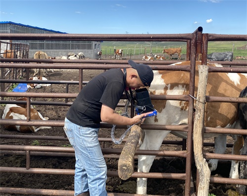
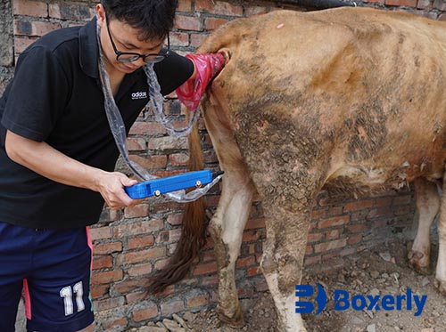
In developed dairy industries such as those in the United States, Germany, and the Netherlands, veterinarians commonly use B-mode ultrasound to track the progress of uterine involution. The scanning is typically done transrectally, allowing clear visualization of the uterine horns, body, and surrounding tissues. This method provides detailed information on wall thickness, fluid accumulation, and intrauterine debris.
Collagen Degradation and Its Role in Uterine Recovery
One of the lesser-known yet scientifically significant aspects of uterine involution is the degradation of collagen in the uterine wall. Collagen provides structural support to the uterus and must be broken down during postpartum regression to enable tissue remodeling and uterine shrinkage.
During pregnancy, the uterus expands to accommodate the growing fetus, which involves a buildup of collagen-rich extracellular matrix. After delivery, enzymatic activity—especially matrix metalloproteinases (MMPs)—breaks down these collagen fibers. A by-product of collagen degradation is hydroxyproline, a unique amino acid that can be detected in the bloodstream. Its presence in serum has been studied internationally, notably in Brazil and Japan, as a marker for tissue remodeling.
Ultrasound does not directly visualize collagen, but it can indirectly indicate the progress of collagen degradation by monitoring uterine wall thickness and tissue echogenicity. A gradually thinning uterine wall combined with reduced internal fluid is a positive sign of collagen breakdown and uterine recovery.
In some cows, however, the collagen degradation process is delayed due to metabolic disorders, nutritional deficiencies, or inflammation. This delay is often seen on ultrasound as a persistently thick uterine wall and excess intrauterine content, signaling incomplete involution and potential subclinical endometritis.
Retained Placenta: A Major Barrier to Recovery
Retained placenta (RP) is defined as the failure to expel fetal membranes within 12 hours after calving. It’s a common problem globally, with reported incidence rates ranging from 10% to 25% and even higher in poorly managed herds. In many cases, RP sets the stage for prolonged uterine inflammation, microbial infection, and delayed uterine involution.
In high-producing dairy cows, RP is often linked to weakened uterine contractions, immune suppression around parturition, or mineral imbalances (notably selenium and vitamin E). Foreign veterinary research, including studies from Canada and Sweden, shows that cows with RP tend to take significantly longer to resume normal estrous cycles and exhibit lower fertility in subsequent inseminations.
With Veterinary ultrasound, RP can be diagnosed within hours post-calving by observing echogenic placental tissue still attached to the uterine lining. Over time, the retained membranes can become necrotic and infected, leading to pyometra (uterine pus accumulation) or metritis. Sonographic indicators include increased uterine diameter, thickened walls, fluid with heterogeneous echotexture, and possibly gas accumulation.
Clinical Signs and Ultrasound Interpretation
Clinically, cows with RP may initially show no systemic signs. However, as decomposition of the placenta sets in, they develop fever, foul-smelling discharge, depression, and reduced feed intake. Ultrasonography provides crucial early warning signs before these clinical symptoms appear.
By scanning cows on days 3, 7, and 14 post-calving, veterinarians can track the uterus’s size reduction, detect any abnormal fluid accumulation, and identify retained tissue. A uterus that does not shrink consistently or shows persistent fluid pockets is often associated with RP or other inflammatory complications.
Farmers and veterinarians in countries like Australia and New Zealand have embraced ultrasound as a standard postpartum tool, allowing for early interventions such as uterine flushing, antibiotic therapy, or manual removal under veterinary guidance.
Comparative Impact on Fertility
Both collagen degradation delay and RP result in prolonged uterine recovery, which has downstream effects on reproductive performance. Studies from European and North American research institutes have found that cows with suboptimal uterine involution are more likely to:
Exhibit delayed estrus cycles
Require multiple inseminations before conception
Experience reduced milk yield due to systemic inflammation
Be culled early due to infertility
Ultrasound-based monitoring not only improves diagnostic accuracy but also enables targeted treatment. For instance, cows with delayed collagen remodeling may benefit from dietary adjustments (e.g., increased protein or micronutrient supplementation) and hormone treatments to stimulate uterine activity. Cows with RP may require prompt antimicrobial therapy to prevent systemic infection.
Ultrasound-Guided Strategies to Improve Outcomes
Veterinary ultrasound has shifted from being a diagnostic-only tool to a proactive management device. On progressive farms, real-time sonography is part of routine postpartum checks and herd reproductive health programs. The benefits of this approach include:
Early detection of complications before clinical symptoms arise
Monitoring treatment effectiveness over time
Reduction in antibiotic misuse through targeted therapy
Shorter calving-to-conception intervals
Improved overall reproductive efficiency and profitability
Conclusion
Uterine recovery is a delicate yet vital process in the postpartum period of dairy cows. Collagen degradation and retained placenta are two major biological and clinical challenges that interfere with this recovery. Veterinary ultrasound allows farmers and veterinarians to visualize and quantify these processes in real time, improving decision-making and reproductive outcomes.
As more dairy operations globally adopt advanced herd management practices, integrating routine ultrasound into postpartum care will become increasingly standard. By doing so, farmers can reduce economic losses associated with infertility, ensure animal well-being, and build more sustainable dairy systems.
Reference Source:
Sheldon, I. M., et al. (2019). Postpartum uterine health in cattle. Reproduction in Domestic Animals, 54(S3), 35–44.
LeBlanc, S. J. (2020). Transition cow health and management. Veterinary Clinics of North America: Food Animal Practice, 36(2), 265–284.
Drillich, M., et al. (2021). Diagnosis and treatment of retained fetal membranes in dairy cows. Veterinary Microbiology, 251, 108907.

