In modern livestock farming, efficient reproduction is essential for profitability and herd health. A key component of reproductive efficiency in female animals is timely and complete uterine involution—the process by which the uterus returns to its pre-pregnant state following parturition. Traditionally, evaluating uterine recovery relied on physical examination or clinical signs, both of which provide limited accuracy. In recent years, however, Veterinary ultrasound technology has revolutionized how we monitor post-partum changes in reproductive organs, especially in cattle, sheep, and horses. While studies on uterine involution using ultrasound exist for large animals, such as cows and mares, the application of this technique in pigs remains largely unexplored. This article discusses how veterinary ultrasound is used globally to assess uterine involution and why this technology holds promise for swine as well.

Understanding Uterine Involution
Uterine involution is a dynamic process involving the reduction of uterine size, the clearing of lochia (postpartum discharge), and the regeneration of the endometrial lining. The completion of involution is vital for the female’s readiness to conceive again, making its accurate assessment critical in reproductive management. Incomplete or delayed involution is often associated with uterine infections, reduced fertility, and economic losses.
In bovine practice, for example, involution generally concludes by 30–40 days postpartum, though this varies by breed, parity, and health status. During this period, changes in uterine diameter, echogenicity of fluids, and the presence or absence of endometrial edema can all be monitored through veterinary ultrasound.
Ultrasound Technology and Technique
Veterinary ultrasound—particularly B-mode imaging—is a non-invasive diagnostic tool that uses high-frequency sound waves to generate real-time images of soft tissues. For assessing uterine involution, a 5.0 or 7.5 MHz transducer is typically inserted transrectally in large animals like cattle and horses. This allows for high-resolution imaging of the uterus and ovaries.
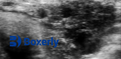
When scanning a non-pregnant uterus, the structure typically appears as a round or slightly oval, hypoechoic (dark) image located just in front of or below the urinary bladder. Several cross-sectional slices can be captured depending on probe placement. In some cases, a small anechoic (very dark) area may appear in the center, usually not exceeding 20–30 mm in diameter. This does not necessarily indicate pathology but should be monitored over time.
The uterus may shift slightly in position depending on bladder fullness. Cross-sectional scans during the post-partum period reveal several identifiable layers within the uterus, including the endometrium, vascular layer, and lochial fluid. As involution progresses, these components undergo clear morphological and echogenic changes.
Global Research on Uterine Involution via Ultrasound
A few notable studies have laid the groundwork for understanding uterine involution through ultrasound. Tian Wertru (1990), Izaike (1989), and Saniont (1994) all conducted significant research in dairy cows using B-mode ultrasound. Their findings confirmed that the uterine horns and body could be measured accurately, and changes in the endometrial and myometrial layers could be observed in real time.
In these studies, post-partum lochia appeared as hyperechoic (bright) or mixed echogenic content within the uterine lumen, gradually diminishing over time. The diameter of the uterine body and horns decreased consistently over the post-partum period, with nearly complete regression observed by the sixth week.
Additionally, these studies demonstrated that ovarian structures such as corpora lutea and follicles could also be monitored. Follicles, which appear as anechoic round areas, could be visualized as small as 2–3 mm using a 5.0 MHz probe. Corpora lutea, by contrast, presented as weakly echogenic areas distinguishable from surrounding ovarian tissue, with subtle changes based on stage—e.g., corpus hemorrhagicum post-ovulation or a mature corpus luteum during luteal phases.
Why Pigs Present a Unique Opportunity
Despite the valuable insights provided by ultrasound studies in ruminants and equines, surprisingly little has been published on monitoring uterine involution in pigs using this technology. This absence is especially notable given the economic importance of swine reproduction worldwide.
Compared to cows, pigs have a more complex uterine structure, including long and coiled uterine horns. However, the application of a transabdominal probe—more practical in pigs due to anatomical constraints—offers potential for frequent, non-invasive monitoring. Recent advances in portable ultrasound machines and real-time image processing make it more feasible to explore this avenue.
Monitoring uterine involution in sows could provide clear benefits, such as earlier detection of retained placenta, subclinical metritis, or delayed ovarian rebound. These conditions often go unnoticed until fertility declines. By identifying and addressing such issues earlier, producers could shorten inter-farrowing intervals and improve overall herd fertility.
Veterinary Ultrasound as a Research and Management Tool
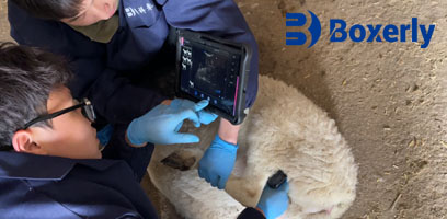
Ultrasound technology is now a standard part of reproductive management in many regions. In the U.S., Canada, and parts of Europe, progressive farms and veterinary clinics routinely scan cows to confirm pregnancy, evaluate ovarian status, and assess uterine recovery. In New Zealand and Australia, ultrasound is also used in sheep operations to optimize lambing schedules and detect reproductive anomalies.
This trend underscores a broader shift in livestock management toward precision farming, where data-driven insights enable better decision-making. Ultrasound fits this model well. It’s fast, repeatable, non-invasive, and delivers immediate visual feedback.
Using Ultrasound in Practice: Key Parameters to Monitor
When evaluating uterine involution via ultrasound, veterinarians and researchers typically assess the following parameters:
Uterine Diameter: A primary indicator of regression. Measured at the body and horns.
Echogenicity of Lochia: Bright or mixed echogenic content suggests early involution. Clear or absent fluid suggests completion.
Wall Thickness and Layer Definition: The reappearance of clearly defined endometrial and myometrial layers indicates progress.
Ovarian Activity: Follicles and corpora lutea signal resumption of cyclicity, a positive sign of reproductive recovery.
Challenges and Limitations
While the benefits are compelling, some challenges remain. In smaller species like pigs or in animals with high fat deposition, ultrasound image quality may be compromised. Operator experience also plays a significant role in accurate interpretation.
Another limitation is the current lack of standardized benchmarks for uterine involution in non-bovine species. This makes comparative analysis difficult and may deter wider adoption in swine or small ruminants.
Nonetheless, ongoing innovation in probe design, software algorithms, and training programs continues to improve usability and accuracy. As interest grows, we can expect more data to emerge that helps define normal versus abnormal patterns in multiple species.
Conclusion
Veterinary ultrasound has emerged as a powerful tool for monitoring uterine involution across several livestock species. While its use in cattle, horses, and sheep is well documented, a promising frontier lies in applying the same technology to pigs—an area still underexplored in both research and practice.
By offering real-time, detailed visualization of uterine and ovarian structures, ultrasound enables earlier interventions, better fertility outcomes, and improved animal welfare. As global livestock systems become more data-driven and welfare-focused, the integration of veterinary ultrasound into routine post-partum evaluations is not just beneficial—it’s inevitable.

