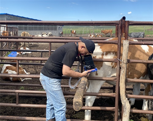
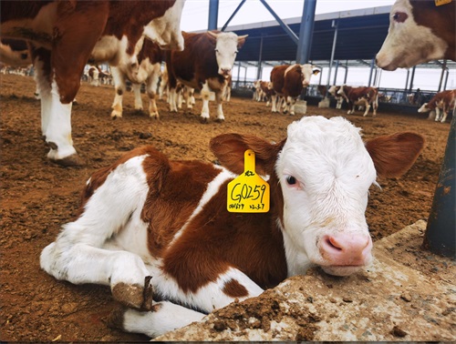
For modern cattle producers and reproductive veterinarians, understanding the physiological changes in a cow's ovary during the estrous cycle is crucial for optimizing artificial insemination (AI), synchronizing estrus, and improving overall fertility outcomes. One of the most powerful tools for this purpose is veterinary ultrasonography—a non-invasive technique that allows us to visualize the ovaries in real time. In the pre-estrus stage, ultrasonography offers valuable information about follicular dynamics, the status of the corpus luteum (CL), and ovulatory readiness.

In this article, we’ll explore how Veterinary ultrasound is used to interpret ovarian changes in cows during the pre-estrus period, supported by real-world images, scientific understanding, and international practices.
Understanding the Pre-Estrus Stage in the Cow
The bovine estrous cycle typically lasts about 21 days and is divided into four stages: proestrus, estrus, metestrus, and diestrus. The pre-estrus phase—also known as proestrus—is the transitional period leading up to visible signs of estrus and ovulation. During this time, the dominant follicle grows under the influence of follicle-stimulating hormone (FSH) and luteinizing hormone (LH), while the corpus luteum from the previous cycle regresses.
From a management standpoint, this is the critical window to monitor because ovulation is imminent. In fixed-time AI programs, knowing exactly when a cow is entering this phase is vital for success.
The Role of Veterinary Ultrasound
Veterinary ultrasound—especially real-time B-mode ultrasonography using 5–8 MHz transrectal probes—is the most effective method to assess ovarian changes during the pre-estrus period. The probe is gently inserted into the cow’s rectum and positioned against the ventral rectal wall to scan the ovaries.
During pre-estrus, ultrasound imaging reveals several characteristic features:
Follicle Identification: Multiple antral follicles (usually 0.3–1.0 cm in diameter) can be observed on both ovaries.
Dominant Follicle Formation: One of these follicles becomes dominant, increasing in size to approximately 1.5–2.5 cm just before ovulation.
Corpus Luteum Regression: The CL from the previous cycle may still be present but often appears smaller, with reduced echogenicity.
Case Example: Interpreting Pre-Estrus Images
Let’s analyze a real ultrasound case to bring theory into practice.
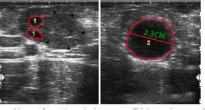
On Day 0 of estrus synchronization protocol, a transrectal ultrasound scan was performed on a multiparous Holstein cow. The images revealed the following:
Left Ovary: Two small follicles measuring 0.5 cm each were visible. No clear CL was detected, although the ovarian stroma appeared slightly heterogeneous, possibly masking a regressing CL.
Right Ovary: A single dominant follicle measuring 2.3 cm was identified. It exhibited a round, anechoic appearance with a smooth wall—typical characteristics of a preovulatory follicle.
This image confirmed the cow was in the late pre-estrus phase, making her an excellent candidate for AI within 24 to 36 hours.
Why the Corpus Luteum Can Be Difficult to Visualize
In some cases, the CL is difficult to distinguish from the surrounding ovarian stroma. This is especially true in the later stages of luteal regression. Because both the CL and stroma are moderately echogenic, they can appear similar on ultrasound. Foreign literature often emphasizes the importance of Doppler ultrasound in these cases, which can help detect luteal blood flow and distinguish functional from regressing CLs.
Twin Ovulations and Double CLs
An interesting phenomenon occasionally observed in pre-estrus cows is twin ovulation—where two dominant follicles develop, one on each ovary, or both on the same ovary. If fertilization occurs, the result may be twin pregnancy.
Ultrasound image A and B from a 28-day post-insemination cow using a 6-MHz linear probe at a depth of 4 cm show the left ovary containing two distinct corpora lutea. This confirms that double ovulation occurred during the pre-estrus phase.
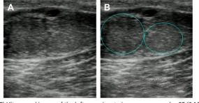
Twin pregnancies are more common in high-producing dairy cows and can lead to both economic opportunities and complications (e.g., increased risk of abortion or dystocia). Recognizing twin CLs on ultrasound early helps veterinarians and farmers plan accordingly.
International Perspective: Use of Ultrasound in Reproductive Management
Across North America and Europe, ultrasound is routinely integrated into reproductive programs. Research from institutions like the University of Wisconsin and Wageningen University in the Netherlands confirms that pre-estrus ultrasonography increases conception rates when paired with fixed-time AI.
In Latin American and Australian beef operations, portable ultrasound devices are becoming increasingly common due to their affordability and ease of use. Operators are trained to evaluate follicle size, CL presence, and ovarian symmetry. The global consensus is clear: ultrasound improves reproductive timing, especially in large herds where visual heat detection is unreliable.
Benefits of Ultrasound During Pre-Estrus
The advantages of using ultrasound during the pre-estrus period are numerous:
Accurate Timing for AI: Identifying dominant follicles allows precise insemination scheduling.
Follicular Monitoring: Detection of abnormal follicular growth, such as follicular cysts.
Estrus Synchronization: Verifying ovarian response to hormonal protocols (e.g., prostaglandin or GnRH injections).
Predictive Value: Dominant follicle presence correlates strongly with imminent ovulation.
Animal Welfare: Non-invasive and repeatable, causing minimal stress to the cow.
Challenges and Limitations
While ultrasonography is a powerful tool, it does require training and experience. Misidentifying structures (e.g., confusing a follicle with a cyst) or failing to detect a CL due to poor contrast are common pitfalls.
Foreign experts often recommend combining ultrasound with hormone profiling—such as measuring progesterone levels—to enhance accuracy. In addition, color Doppler technology is gaining popularity for better differentiation of active vs. inactive CLs.
What Makes a Good Pre-Estrus Ultrasound Technician?
From discussions with veterinary schools and AI service providers globally, a skilled ultrasound technician during pre-estrus monitoring typically exhibits the following qualities:
Deep understanding of bovine ovarian physiology.
Familiarity with probe positioning and angle adjustment.
Ability to interpret subtle echogenic differences in ovarian tissue.
Knowledge of the hormonal cycle and synchronization protocols.
Conclusion: Integrating Ultrasonography into Reproductive Strategies
Veterinary ultrasound has revolutionized the way we monitor ovarian activity in cows, especially during the crucial pre-estrus phase. By visualizing follicular development and CL status in real time, we can optimize AI timing, improve pregnancy rates, and enhance herd reproductive performance.
As portable, user-friendly ultrasound machines become more accessible, even small-scale farmers can benefit from integrating this technology into their herd management practices. In the future, combining real-time imaging with automated estrus detection and hormone monitoring may create a fully integrated reproductive management system.
Whether you are managing a 50-cow dairy or a 1,000-head beef operation, mastering the art of pre-estrus ovarian imaging is a step toward greater reproductive success.

