As a responsible pet owner or veterinary professional, ensuring the health of a pregnant dog is a matter of critical importance. Among the various diagnostic tools available today, Veterinary ultrasound has emerged as one of the most effective, non-invasive, and widely accepted methods for evaluating reproductive health. This includes detecting abnormal uterine fluid accumulation and monitoring fetal development throughout the gestation period.
In this article, we will explore how veterinary ultrasound is used to identify uterine abnormalities and assess fetal health in pregnant bitches. Drawing on both practical experiences and international veterinary standards, we will also discuss key precautions that ensure accurate diagnosis and effective monitoring.

Understanding the Role of Veterinary Ultrasound in Canine Reproduction
Veterinary ultrasound, particularly B-mode (brightness mode) imaging, offers real-time, cross-sectional visualization of internal organs. In reproductive evaluations, it allows veterinarians to observe the uterus, ovaries, and developing fetuses with exceptional clarity. The primary benefit is its non-invasive nature, which makes it ideal for frequent monitoring without exposing the dog or fetuses to ionizing radiation.
In canine reproduction, ultrasound can be used as early as 20 to 25 days after ovulation to confirm pregnancy, detect fetal heartbeats, and estimate the number of fetuses. Beyond that, it is invaluable in identifying pathological conditions such as pyometra (uterine infection), fetal death, resorption, and abnormal fluid accumulations.
Detecting Abnormal Uterine Fluid Accumulation
In clinical practice, one of the most common uterine abnormalities seen via ultrasound is the presence of abnormal intrauterine fluid. This could be due to various conditions such as hydrometra, mucometra, or pyometra. Early detection is crucial to prevent complications that could jeopardize the dog’s health or pregnancy outcome.
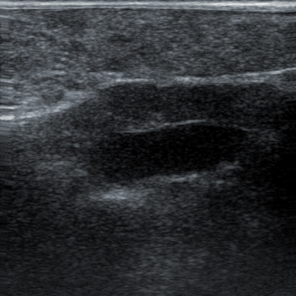
When scanning in parallel with the uterine body or horns, abnormal fluid typically appears as an anechoic (dark, echo-free) tubular area with uniform internal texture. These fluid-filled structures may present as elongated, round, or irregularly shaped zones depending on the scanning angle and severity of the condition. Their walls are generally visible and more prominent compared to early pregnancy sacs. In cross-sectional or transverse views, these structures often appear circular or lobulated and should not be confused with fetal sacs.
A key diagnostic feature is the absence of fetal echoic patterns within these fluid zones. Unlike normal pregnancy sacs that display small echogenic dots (early fetal poles or placental tissues), abnormal fluid accumulations are homogeneously dark. There is typically no floating material or fetal reflections within the lumen. Additionally, the surrounding intestinal loops, containing gas and fluid, may appear adjacent to these structures but can be differentiated by their peristalsis and layered wall appearance.
Internationally, veterinary protocols emphasize the importance of differentiating between uterine fluid build-up and early gestational sacs. Misdiagnosis can lead to inappropriate treatment or missed opportunity for early intervention in cases such as closed pyometra, which requires immediate surgical management.

Ultrasound Features of Early Pregnancy in Dogs
To distinguish abnormal fluid from early pregnancy, it is essential to understand the normal sonographic appearance of pregnant dogs. During the first 24 to 40 days of gestation, ultrasound is the preferred method for monitoring development. Pregnancy sacs appear as small, round, anechoic areas with a thin echogenic rim (representing chorionic tissue). Over time, these sacs become more clearly defined, and fetal poles, placental zones, and eventually heartbeats become visible.
Early in pregnancy, amniotic fluid occupies a larger portion of the uterine cavity, and the fetus itself may still be quite small. This makes precise measurements challenging and prone to error, especially when there are overlaps or deformation due to sac crowding after day 35. At this stage, it becomes increasingly important to rely on fetal landmarks—such as spine and head reflections—to differentiate fetuses from fluid anomalies.
Measuring Fetal Development: Best Practices and Pitfalls
One of the primary uses of ultrasound in canine obstetrics is to estimate fetal age, body length, and viability. However, practitioners must be aware of certain limitations and standard precautions to avoid misinterpretation.
During the earlier stages of pregnancy (24 to 35 days), fetal growth is inconsistent. While gestational sacs grow rapidly, the embryo develops more slowly. This is attributed to the relatively large volume of amniotic fluid and the ongoing formation of fetal membranes. Measurements such as gestational sac diameter, crown-rump length (CRL), and head diameter are useful, but their accuracy depends on clear identification of fetal landmarks and a steady probe hand.
After day 35, sac crowding becomes more pronounced, and gestational sacs may change shape due to uterine compression. This can lead to difficulty in obtaining consistent measurements and cause over- or underestimation of fetal age. Thus, veterinarians are advised to take multiple measurements and average the results to improve accuracy.
It’s also important to recognize the presence of placental tissue around the fetus, which may interfere with the visualization of clear fetal boundaries. In these cases, applying slight probe pressure and using multiple scanning planes (longitudinal and transverse) can help delineate anatomical details more effectively.
Physiological Considerations: Understanding Pregnancy Dynamics
Canine pregnancy is a dynamic process involving the coordinated growth of embryos, development of placental attachments, and progressive expansion of the uterus. Fertilization is only the starting point; successful implantation and fetal viability depend on maternal hormonal support and uterine health.
Ultrasound plays a crucial role not only in morphological assessment but also in evaluating physiological parameters such as fetal heart rate and placental structure. Abnormalities such as reduced fetal movement, bradycardia, or placental detachment are red flags indicating fetal distress and the need for immediate veterinary intervention.
Veterinarians trained in ultrasonography are expected to have a thorough understanding of uterine anatomy, reproductive physiology, and common pathologies. According to international guidelines, practitioners must correlate sonographic findings with clinical signs and laboratory results to arrive at a reliable diagnosis.
Best Practices in Using Veterinary Ultrasound on Pregnant Dogs
To ensure reliable outcomes and minimize error during ultrasonographic examination, several best practices should be followed:
Patient Preparation:
Ensure the dog is calm and lying in dorsal or lateral recumbency.
Shave or clip the abdominal hair and apply ultrasound gel to avoid air interference.
Fasting before scanning can reduce intestinal gas, which may obstruct imaging.
Probe Handling:
Use a high-frequency (5–10 MHz) linear or convex transducer for optimal resolution.
Scan both longitudinal and transverse planes for full uterine coverage.
Maintain gentle, consistent pressure to avoid causing discomfort.
Image Interpretation:
Familiarize yourself with normal anatomical variants and gestational timelines.
Record multiple views and measurements for comparison.
Monitor fetal heart rate and movement regularly as indicators of viability.
Documentation and Follow-up:
Save and label images for each fetus or uterine segment.
Schedule rechecks during critical gestational stages (e.g., around day 25, 35, and 45).
Educate pet owners on expected outcomes, signs of complications, and post-birth care.
Global Perspectives and the Growing Importance of Canine Obstetric Imaging
Internationally, there is a growing emphasis on the role of veterinary ultrasound in small animal practice. Organizations such as the American College of Veterinary Radiology (ACVR) and European College of Veterinary Diagnostic Imaging (ECVDI) have developed detailed training and certification pathways to ensure practitioners can accurately diagnose reproductive conditions.
In countries like the United States, Canada, Germany, and Australia, ultrasound is often the first line of diagnostic imaging for pregnant pets, thanks to its safety and diagnostic value. Moreover, with the rise in pet ownership and breeding programs, there is a parallel demand for accurate, timely prenatal care—making ultrasonography a standard part of canine reproductive medicine.
Conclusion
Veterinary ultrasound has transformed the way we approach pregnancy monitoring and uterine health in dogs. From identifying abnormal fluid accumulation to tracking fetal growth and viability, this technology provides real-time, invaluable insight for veterinarians and breeders alike. However, its accuracy and usefulness depend on proper technique, anatomical knowledge, and interpretation skills.
By following best practices and staying updated with international veterinary standards, practitioners can ensure better reproductive outcomes, improve maternal care, and support healthy litters. For dog owners, regular prenatal ultrasound exams not only bring peace of mind but also offer a tangible connection to the new lives developing inside their beloved pet.
link: https://www.bxlimage.com/nw/1213.html
tags:


