Infectious arthritis is a serious condition in cattle, often leading to lameness, reduced productivity, and significant economic losses. As a livestock farmer, I’ve encountered cases where early and accurate diagnosis is crucial to managing the health of the herd. Traditional diagnostic methods have their limitations, especially in early-stage infections. In recent years, ultrasound imaging has emerged as a reliable and non-invasive technique to detect joint disorders, including infectious arthritis. This article explores how bovine ultrasound is used to evaluate joint infections and highlights findings from studies comparing ultrasound with traditional diagnostic techniques.
")
Understanding Infectious Arthritis in Cattle
Infectious arthritis in cattle commonly develops as a secondary complication of conditions like sole ulcers, interdigital dermatitis, and foot rot. The infection can invade the joint capsule, leading to inflammation, fluid accumulation, and tissue damage. If not diagnosed early, it may result in irreversible joint damage and necessitate culling. Thus, early detection is vital not only for treatment success but also for making informed decisions about animal welfare and herd management.
Limitations of Traditional Diagnosis
Traditionally, diagnosing joint inflammation in cattle involves:
Physical and lameness examinations: Observing gait and joint swelling.
Joint aspiration: Collecting synovial fluid with a needle.
Radiography (X-rays): Visualizing bone changes.
While these methods are valuable, they have several drawbacks. In early-stage infections, joint fluid may not be easily obtained through aspiration, and radiographs might not show clear abnormalities. Moreover, physical signs like swelling or lameness can be subtle or mistaken for other issues. As a result, some cases may go undetected until they become more severe.
The Role of Ultrasound in Diagnosing Bovine Arthritis
Ultrasound offers a non-invasive, real-time imaging method that provides detailed visualization of soft tissues, including joint capsules, tendons, ligaments, and synovial fluid. This makes it particularly effective for detecting inflammatory conditions like infectious arthritis before major structural changes occur.
In my experience, and as supported by veterinary studies, ultrasound can identify abnormalities that would otherwise be missed. For instance, in a study involving ten cattle with confirmed infectious arthritis and limb amputations, ultrasonographic findings were compared with conventional methods and post-mortem analysis. The results confirmed that ultrasound reliably detected signs of joint infection.
Key Ultrasonographic Findings in Infectious Arthritis
The ultrasound examination revealed several consistent indicators across the infected limbs:
Enlarged joint capsule: A hallmark sign, appearing as an anechoic (dark) area on the image, indicating fluid accumulation.
Joint capsule measurements:
Dorsal side: average thickness of 7.9 mm (range: 5.8–12.3 mm)
Palmar/plantar side: average thickness of 13.8 mm (range: 10.8–16.4 mm)
Other abnormalities observed on palmar/plantar images:
Presence of hoof abscesses
Torn deep digital flexor tendon
Osteomyelitis of the distal sesamoid bone
Excess synovial fluid in the tendon sheath
These findings were consistent with joint infections confirmed through necropsy. Importantly, such detailed visualization is not possible with radiography or clinical examination alone.
Advantages of Ultrasound Over Conventional Methods
From a farmer’s point of view, the advantages of using ultrasound include:
Early detection: Identifies inflammation before radiographic signs appear.
Real-time assessment: Allows on-the-spot evaluation and decision-making.
Non-invasive: Reduces stress and injury risk compared to joint aspiration.
More precise treatment planning: Guides targeted therapy or surgical intervention.
Additionally, ultrasound does not expose animals or handlers to ionizing radiation, making it safer for routine use.
Clinical Implications and Decision-Making
Diagnosing infectious arthritis early means veterinarians can begin treatment promptly—such as administering antibiotics or anti-inflammatory medications—and monitor progress. In severe cases where prognosis is poor, early diagnosis via ultrasound can support timely decisions regarding euthanasia or culling, reducing unnecessary suffering.
For farms where lameness is a recurring issue, incorporating ultrasound into routine diagnostics can improve herd health outcomes and lower long-term treatment costs.
Integration into Farm Practice
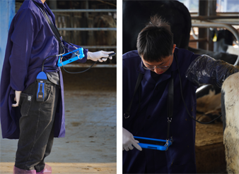
While ultrasound equipment may seem complex, portable and ruggedized Veterinary ultrasound units have made it more accessible for use in field conditions. With basic training and the guidance of a veterinarian, farm personnel can assist in identifying animals needing further assessment.
Moreover, as ultrasound technology advances, images become clearer and interpretation more intuitive, making the process easier for both vets and farmers.
")
Conclusion
Ultrasound has proven to be a powerful tool in detecting infectious arthritis in cattle. It provides accurate, early diagnosis, allowing for timely and effective treatment. By visualizing fluid buildup, tendon damage, and bone infection within the joint, it fills a crucial gap left by traditional diagnostic methods.
As a livestock producer, adopting ultrasound as part of the health management toolkit has helped me make faster, better-informed decisions for my herd. Whether for identifying joint issues, reproductive monitoring, or musculoskeletal evaluation, ultrasound is becoming an indispensable technology on modern farms.
References
Braun, U., et al. (2007). Ultrasonographic examination of the bovine digit. Veterinary Journal.
Kofler, J. (2013). Diagnostic imaging in lameness evaluation in cattle. Veterinary Clinics of North America: Food Animal Practice.
Desrochers, A. (2004). Musculoskeletal disorders of the bovine digit. Veterinary Clinics of North America: Food Animal Practice.
link: https://www.bxlimage.com/nw/1205.html
tags: