Ultrasound imaging, or ultrasonography, is one of the most widely used diagnostic tools in veterinary medicine, especially for evaluating soft tissues in large animals. Its non-invasive nature, real-time feedback, and relatively low cost make it a critical asset in animal health management, from reproductive checks to musculoskeletal evaluations.
")
Basic Principles of Ultrasound
Ultrasound works on the principle of sound wave reflection. High-frequency sound waves—typically between 1.5 to 18 megahertz (MHz)—are emitted into the animal’s body using a device called a transducer. These waves travel through tissues at different speeds, depending on tissue density. When they encounter a boundary between different tissues (such as fluid and muscle or muscle and bone), a portion of the wave reflects back to the transducer as an echo.
The transducer acts both as a transmitter and receiver. It converts the returning echoes into electrical signals, which are then processed by the ultrasound machine to generate an image. This image represents a real-time, cross-sectional view of the internal anatomy.
Image Formation and Modes
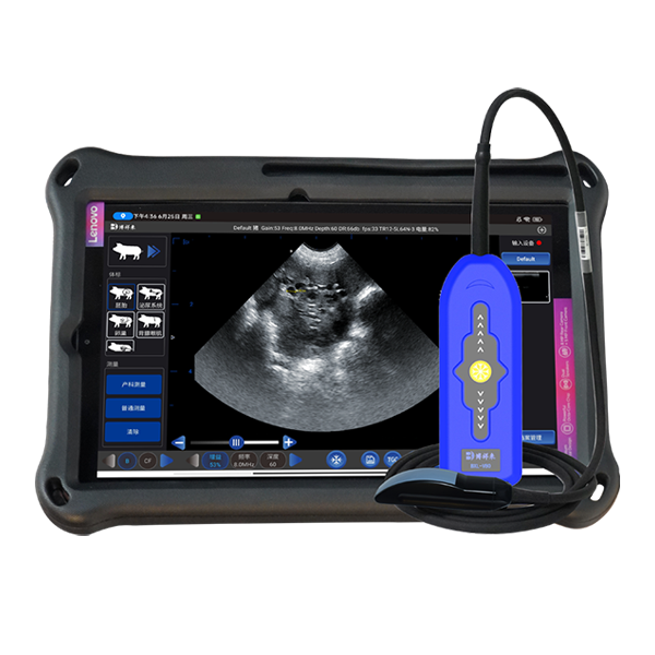
The most commonly used imaging mode in veterinary practice is B-mode, or brightness mode. In B-mode, echoes are displayed as varying shades of gray. The brighter the echo, the more solid or dense the tissue. This mode is ideal for general anatomical imaging.
Other modes include:
M-mode (Motion mode): Primarily used in cardiac imaging to assess the movement of heart structures.
Doppler mode: Evaluates blood flow velocity and direction using the Doppler effect, where sound frequency changes with movement.
Color Doppler: Adds color overlays to blood flow images to indicate direction and speed.
Power Doppler: Enhances sensitivity to low-velocity blood flow.
The Role of Gel
A transmission gel is applied to the skin before scanning. This eliminates air gaps between the transducer and the skin surface, which could otherwise prevent sound wave transmission. Air is a poor conductor of ultrasound, while gel ensures proper acoustic coupling for accurate imaging.
Tissue Interaction and Echo Patterns
Echoes are created when sound waves transition between tissues of different acoustic impedance. For example, when waves hit the boundary between muscle and fluid, some of the waves reflect while others pass through. The greater the difference in density, the stronger the echo.
This principle also explains why ultrasound does not image well through gas or bone. Gas causes total reflection, and bone absorbs the ultrasound, producing "shadowing" effects behind these structures. Therefore, imaging organs behind gas-filled intestines or dense bones can be challenging.
")
Depth and Frequency Considerations
Ultrasound penetration depth is inversely related to frequency. Low-frequency transducers (e.g., 2–5 MHz) penetrate deeper but have lower resolution, making them suitable for large animals. High-frequency transducers (e.g., 7.5–15 MHz) offer better resolution but shallow penetration, ideal for superficial structures.
In most cases, ultrasound can effectively image tissues up to 30 cm deep. However, beyond this depth, echoes may become too weak, or noise may obscure the image due to energy dispersion.
Common Applications in Large Animal Practice
Reproductive Assessments
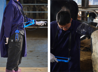
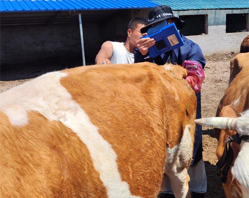
Ultrasound is extensively used for reproductive monitoring in cattle, horses, sheep, and goats. Transrectal probes allow veterinarians to assess ovarian structures, diagnose pregnancy, estimate fetal age, and detect reproductive abnormalities with high precision.
Musculoskeletal Evaluations
Tendon and ligament injuries in horses are often diagnosed and monitored using high-frequency linear probes. Changes in tendon fiber alignment, echogenicity, and cross-sectional area provide information about injury severity and healing.
Abdominal Imaging
In large ruminants, abdominal ultrasound assists in diagnosing gastrointestinal obstructions, liver abscesses, and peritonitis. However, bowel gas often limits visibility, requiring skilled manipulation to acquire interpretable images.
Thoracic Imaging
Ultrasound can visualize the pleural surface, fluid accumulation (pleural effusion), or abscesses in the chest cavity. However, the lungs themselves are usually inaccessible due to air interference.
")
Echocardiography
Echocardiography is a specialized form of ultrasonography used to evaluate heart anatomy and function. Using M-mode and 2D imaging, veterinarians can assess valve motion, chamber size, wall thickness, and contractility. Doppler imaging further quantifies blood flow abnormalities like regurgitation or stenosis.
Some systems now offer 3D echocardiography and automated cardiac measurements, improving diagnostic accuracy in both large and small animals.
Ultrasound-Guided Procedures
Ultrasound guidance enhances the safety and accuracy of needle-based procedures. This includes:
Biopsy of internal organs (liver, kidney, lymph nodes)
Fine-needle aspiration of masses
Fluid drainage from abscesses or cysts
These techniques often eliminate the need for exploratory surgery and can be performed under sedation rather than general anesthesia, especially in livestock.
Limitations of Ultrasound
While highly valuable, ultrasound has certain limitations:
It cannot penetrate air-filled or calcified structures.
Image quality deteriorates with depth and noise.
It requires substantial operator training and anatomical knowledge.
It is less effective for comprehensive evaluation of bone interiors or airways.
")
Advances in Veterinary Ultrasound
Modern ultrasound machines have enhanced image resolution, real-time video recording, and digital storage (e.g., DICOM format). Software updates allow tissue characterization, automated measurements, and even contrast-enhanced ultrasonography. In contrast-enhanced studies, microbubble agents are injected intravenously to assess tissue perfusion and vascularity.
These technological improvements expand the role of ultrasound from mere diagnostic imaging to interventional and prognostic applications in veterinary practice.
References
Merck Veterinary Manual. Ultrasonography in Large Animals. Retrieved from: https://www.merckvetmanual.com/clinical-pathology-and-procedures/diagnostic-imaging/ultrasonography-in-large-animals
American College of Veterinary Radiology (ACVR). Ultrasound Imaging. https://acvr.org
International Veterinary Information Service (IVIS). Veterinary Ultrasonography. https://www.ivis.org
link: https://www.bxlimage.com/nw/1201.html
tags: