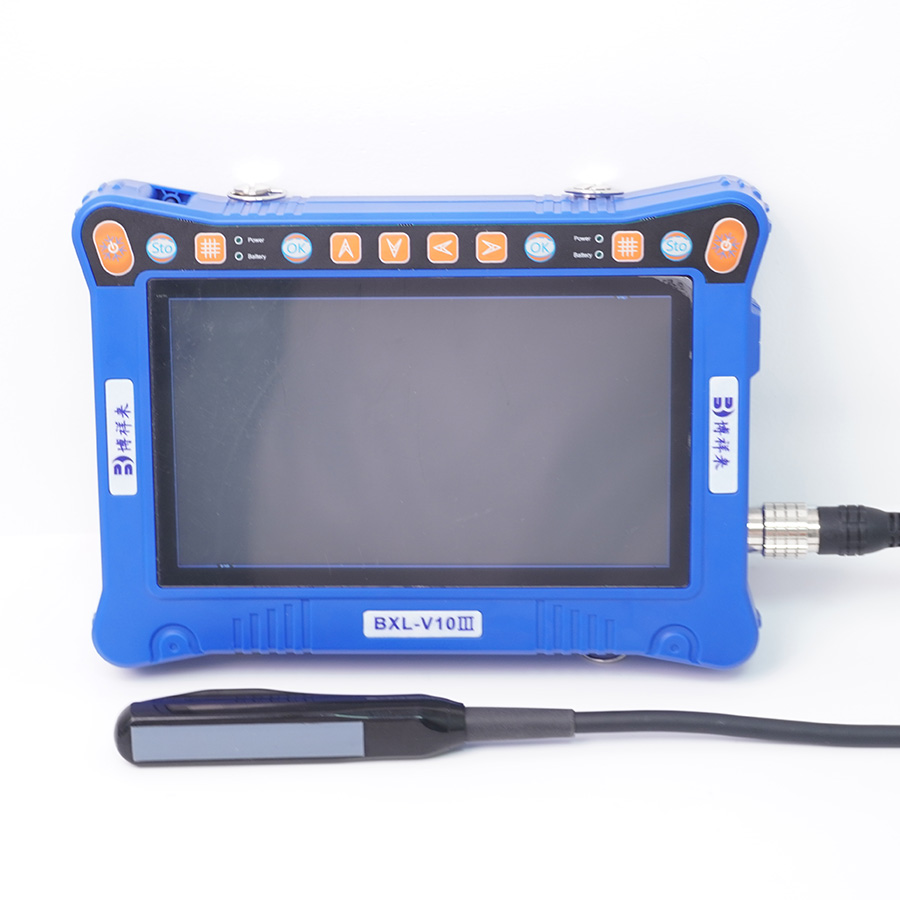
Animal B-ultrasound instrument rectal exploration method: Rectal exploration is mostly performed with a 5.0MHz linear array probe or a higher frequency rectal probe. Boxianglai BXL-V10Ⅲ veterinary B-ultrasound is equipped with a 5.0MHz linear array probe, but its handle is very short, so a plastic jacket of about 30 cm is required for rectal exploration to allow it to reach deep into the rectum and rotate the probe.

During the exploration, the sheep is naturally standing and restrained, and the feces in the rectum are first discharged. The long-handled probe is disinfected and moistened in disinfectant and then inserted into the rectum with the exploration surface facing down, and scanned section by section. It is sent to the entrance of the pelvic cavity, and then carefully scanned downward and tilted 45° to both sides. After scanning the appropriate image, stabilize the probe, freeze the image first, and then take a photo, or store and print it.
Animal B-ultrasound instrument rectal sonogram performance: The first section after the probe enters the rectum can show the vaginal structure and pubic symphysis below the rectum. Because the sound beam cannot penetrate the pubic bone, an acoustic shadow appears below the pubic bone. When there is no fluid accumulation in the vagina, the strong echo light bands of the upper and lower serous membranes can be displayed, with the muscle layer and mucosa layer of medium-intensity echo in the middle. Because the difference in acoustic impedance values is not large, the hierarchical structure is not clear, but the double-layer thickness between the serous membrane light bands can be measured. If there is fluid accumulation in the vagina, the double-layer mucosa can be displayed, and a liquid dark zone can be displayed between the mucosal layers. The vestibular glands of sheep can be clearly displayed deep in the vagina, showing a solid dark area mixed with echo light spots, which is approximately circular and about 1 × 1.5 cm in size.
The second section of the sheep B-ultrasound probe that continues to move forward can show the cervix, uterine body, bladder neck and the position of the anterior pelvic opening. The bladder neck is an inverted pear-shaped liquid dark area, and the bladder wall can be clearly displayed. The pubic symphysis at the anterior opening of the pelvic cavity is a strong echo band, and the acoustic shadow of the posterior band can clearly distinguish the boundary between the abdominal cavity and the pelvic cavity.
When the B-ultrasound probe of sheep enters the abdominal cavity from the pelvic cavity and scans to the left or right at a 45-degree angle, the third section can show abdominal organs such as the bladder, uterine horn, ovary, rumen, and intestinal tube. Increasing the display depth can reveal the strong echo light band with acoustic shadow on the lower abdominal wall. The bladder is a circular liquid dark area. A solid dark area with mixed echo light spots can be clearly seen in the front and lower part of the bladder, which is the echo structure of the uterine horn and ovary. The solid dark area can distinguish the cross-sectional structure of the uterine horn and the oval ovarian structure with a surrounding echo band (formed by the ovarian capsule and the outer fat layer). If it is a pregnant sheep, the liquid dark area and embryonic node formed by the gestational sac will be visible in this solid dark area. If the animal is in estrus, a small circular liquid dark area formed by the follicle or a corpus luteum structure with strong echo will be visible in the ovarian solid dark area. The intestinal tube is often manifested as a cloud-like strong echo light group with acoustic shadow behind it due to gas accumulation. When the examination was performed after full feeding, a strong echo band formed by the blind sac behind the rumen could be seen on the left side.


