")
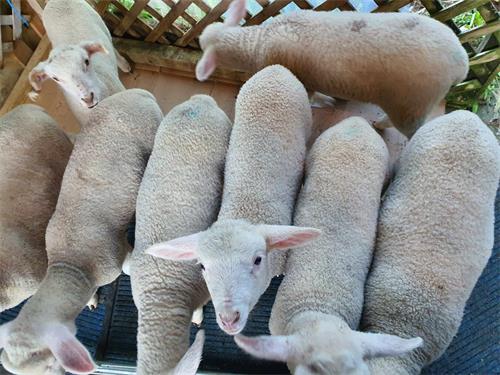
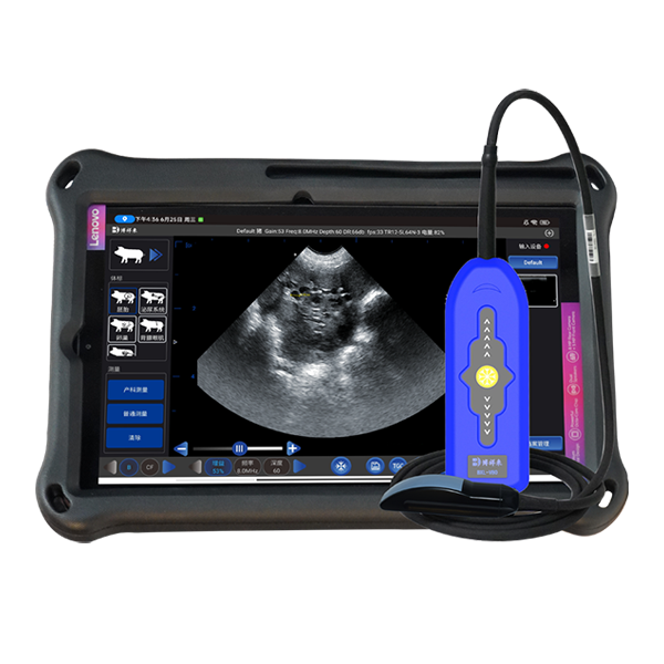
When the ewe has uterine prolapse, a vertically growing object is exposed from the vulva, and there are symptoms such as arched waist and restlessness. The prolapsed uterus is bright red at first, shiny, with many dark red maternal placentas, and is very easy to bleed. After a while, congestion, edema, and mucosal cracking will occur, often with feces, bedding, mud and other dirt. If the uterus is prolapsed for too long, the mucosa will be necrotic, and peritonitis, sepsis, etc. may occur, and systemic symptoms may occur. Use sheep B-ultrasound for auxiliary diagnosis, and then treat and repair after confirmation.
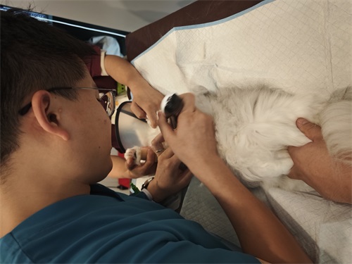
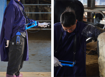
When repairing the prolapsed uterus of the ewe, after thorough cleaning and disinfection, the assistant will lift the uterus to the same height as the vulva with a disinfected basin. The surgeon will hold the tip of the ewe's uterus with his fist and carefully push forward while the ewe is not straining: it can also start from the base of the uterus. The surgeon will use both hands to push inward alternately from both sides of the vulva, pushing part by part, and returning all the prolapsed uterus to the pelvic cavity. During the pushing process, the sheep B-ultrasound should be used for monitoring. In order to prevent the prolapsed uterus from being reset in time, the surgeon can reach into the uterus for correction, and the uterus will be reset under the monitoring of the sheep B-ultrasound.
Prevention and treatment After repair, in order to prevent infection, the surgeon sprinkles penicillin or 20% amoxicillin powder into the uterus during correction, and continuously injects penicillin or amoxicillin intramuscularly for 3-5 days: After repair, in order to prevent the uterus from prolapse again. The ewe can be placed in a low front and high back position, and 1-2 buttonholes are sutured from the upper part to the middle part of the vulva, and the sutures are removed after 3-4 days. After recovery, strengthen feeding management, use sheep B-ultrasound frequently to check its recovery, and give nutritious and juicy green feed to help it recover as soon as possible.
link: https://www.bxlimage.com/nw/634.html
tags: sheep B-ultrasound ultrasound sheep ultrasound