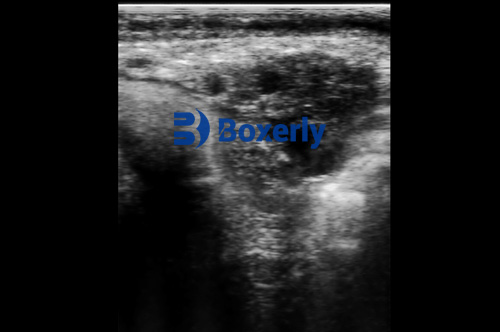
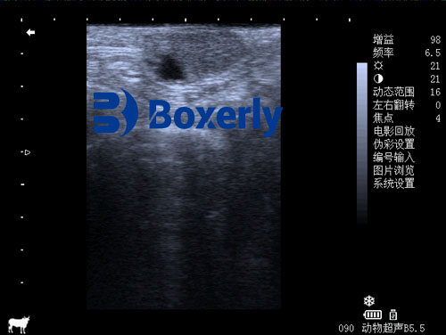
In modern cattle reproduction management, determining the optimal time for breeding is one of the most critical factors in improving conception rates and maximizing reproductive efficiency. For decades, livestock farmers relied primarily on behavioral observations and rectal palpation to estimate estrus and ovulation timing. However, as reproductive biology and veterinary technology have advanced, ultrasonography—especially B-mode ultrasound—has emerged as a highly accurate, non-invasive tool for monitoring ovarian follicular development and predicting the best time for artificial insemination (AI).

This article explores how ultrasonography is used to determine the development of ovarian follicles and the breeding time of cattle. Drawing from both field practice and international research, we will examine the biological processes, the limitations of traditional methods, and how B-mode ultrasound provides a revolutionary solution.
Understanding the Ovarian Cycle in Cattle
Cattle are polyestrous animals, meaning they cycle regularly throughout the year. A typical estrous cycle lasts 18–24 days and is driven by fluctuations in hormones like estrogen, progesterone, and luteinizing hormone (LH). Central to this cycle is the growth and regression of ovarian follicles.
Ovarian follicles develop in waves. In each wave, several follicles begin to grow, but usually only one becomes dominant and eventually ovulates. The timing of this ovulation is critical: if insemination occurs too early or too late, fertilization may not occur, or the embryo may fail to implant properly.
Traditionally, farmers detect estrus by observing standing heat—a behavior where a cow stands still when mounted by another. However, standing estrus in dairy cows is often brief and subtle, sometimes lasting less than 12 hours. Misjudging this narrow window can drastically lower the conception rate.
The Role of Ultrasonography in Follicular Monitoring
Ultrasonography, particularly B-mode (brightness mode) imaging, enables veterinarians to visually monitor the ovaries in real-time. Using a transrectal probe, they can identify and measure the diameter of developing follicles, confirm the presence of a corpus luteum (CL), and detect abnormalities like cysts or inactive ovaries.
When applied consistently, ultrasound can detect:
Follicular wave patterns
Dominant follicle size and status
Signs of imminent ovulation (e.g., thinning follicular walls)
Post-ovulatory changes and CL formation
This information allows farmers and veterinarians to schedule AI more precisely, improving pregnancy success rates and reducing repeat breedings.

Why Not Just Use Rectal Palpation?
In some contexts, rectal palpation still has value—especially where access to ultrasound is limited. For example, in sex-controlled frozen semen applications, palpation can help identify the ovary with a mature follicle so that semen can be injected into the corresponding uterine horn. However, this method is limited by a few key factors:
Palpation cannot confirm follicle rupture – If the fimbriae of the fallopian tube do not relax in time after follicle rupture, the egg may fall into the abdominal cavity rather than the oviduct, reducing fertilization chances.
Physical stress – Touching the follicle adds unnecessary stress to the animal and increases workload, often without meaningful reproductive benefit.
Short estrus duration – Since estrus signs are fleeting (lasting <12 hours), relying on behavioral cues or palpation risks missing the optimal breeding window.
In contrast, ultrasonography provides a clear, repeatable, and quantitative assessment of the reproductive cycle.
Breeding Timing: Synchronizing Biology with Precision
Research has shown that the interval from the onset of standing estrus to ovulation is typically 24–32 hours. Sperm can survive in the reproductive tract for 24–30 hours, while an egg remains viable for only about 10 hours post-ovulation. This means timing AI within a narrow window—ideally around 8–12 hours after the onset of standing estrus—offers the best chance for successful fertilization.
However, in real-life scenarios, standing estrus may be missed altogether. That’s where ultrasound shines. By observing the size and development of dominant follicles, trained professionals can estimate when ovulation is likely to occur and advise on optimal insemination time—even in silent heats or irregular cycles.
Additionally, ultrasound helps detect:
Anovulation – Where no dominant follicle develops
Cystic ovaries – Follicles that grow but do not rupture, leading to infertility
Luteal insufficiency – Where the corpus luteum does not form or function properly after ovulation
Detecting these conditions early helps target interventions and avoid unnecessary insemination costs.

Special Cases: Reproductive Disorders and Infertility
In cases of repeat breeders or cows with unclear estrus behavior, ultrasonography becomes a diagnostic powerhouse. It allows for:
Assessment of ovarian activity in cows with suspected estrus disorders
Differentiation between true anestrus and silent estrus
Monitoring follicular dynamics in response to hormone treatments (e.g., PGF2α or GnRH protocols)
Evaluating the success of synchronization protocols used in timed AI (TAI) programs
For example, if a cow has a normal uterus but lacks follicular growth, it may suggest hormonal imbalance. Alternatively, finding a persistent follicle could explain repeated failure to conceive.
These nuanced insights cannot be gleaned through rectal palpation alone.
International Perspective: Why Ultrasonography is Gaining Ground Globally
In North America, Europe, and Australia, the adoption of veterinary ultrasound in reproduction programs has become mainstream. According to the Journal of Dairy Science, farms that utilize ultrasound for reproductive monitoring see a 15–25% improvement in overall pregnancy rates and a reduction in calving intervals.
European researchers also highlight ultrasound’s role in reducing hormonal use. By precisely timing insemination based on follicular development, fewer hormone-based synchronization treatments are needed, aligning with animal welfare and organic farming standards.
Furthermore, portable ultrasound systems, like the BXL-V50, have made it easier for farm vets to perform high-resolution scans in field conditions. With long battery life, waterproof design, and real-time imaging, such devices empower on-farm decision-making without needing to transport animals to clinics.
Technological Benefits of B-mode Ultrasound in Breeding Management
Non-invasive monitoring of follicular size and morphology
Early detection of ovulation, luteal phase, or reproductive pathologies
Improved AI success rates, especially when using expensive sexed semen
Lower costs through reduced reliance on hormone synchronization
Real-time feedback during breeding protocols and timed insemination programs
These advantages make B-mode ultrasonography not just a tool—but a strategic asset—in reproductive herd management.
Integrating Ultrasound into a Breeding Strategy
An effective reproductive program using ultrasound may include the following steps:
Baseline examination of all breeding-age cows for reproductive tract health
Regular follicular monitoring every 48–72 hours to track follicular waves
Timed AI based on dominant follicle size (typically >10 mm) and wall thinning
Post-breeding examination at 24–48 hours to confirm ovulation and early CL development
Pregnancy confirmation via ultrasound as early as 25–30 days post-breeding
Such a structured approach not only improves conception rates but also allows better culling and management decisions.
Conclusion: A New Era in Cattle Breeding Efficiency
In today's competitive agricultural landscape, every missed breeding cycle represents lost time and money. The use of B-mode ultrasonography to determine ovarian follicular development and breeding time in cattle is a major leap forward in reproductive management.
By replacing guesswork with real-time data, farmers and veterinarians can ensure more timely, efficient, and successful inseminations. Whether dealing with high-value animals, managing large herds, or using costly sexed semen, this technology reduces risks and increases returns.
As access to affordable, rugged, and portable ultrasound systems improves worldwide, such as with the BXL-V50, the integration of ultrasound into daily herd management will only grow. For cattle producers seeking precision, efficiency, and results, B-mode ultrasound is no longer optional—it’s essential.
References
Whitaker, D. A., & Smith, E. (2021). Veterinary Ultrasonography in Food-Producing Animals. Journal of Veterinary Imaging.
Fricke, P. M., & Giordano, J. O. (2020). Reproductive physiology and management of dairy cows using ultrasonography. Journal of Dairy Science, 103(10), 9775–9790.
Beef Cattle Institute. (2023). “Use of Ultrasound for Growth and Reproduction in Cattle.” https://www.beefcattleinstitute.org/ultrasound-growth
Houghton, P. L., & Turlington, L. M. (2022). Using Ultrasound in Beef Cattle Reproduction. Iowa State Extension. https://www.extension.iastate.edu/livestock/ultrasound-reproduction
link: https://www.bxlimage.com/nw/230.html
tags: B-ultrasound of cattle B-ultrasound


