Subfertility in cows is a growing concern among farmers, especially in open farm systems where natural breeding and seasonal calving are preferred. With fewer cows conceiving at expected intervals, the pressure on veterinary diagnostics has never been higher. One of the most transformative tools in addressing this challenge is portable ultrasound. It's not just for confirming pregnancy anymore—it's now a key player in understanding the deeper story of uterine structure and its impact on fertility.
")
The Challenge of Subfertility in Open Farm Systems
Subfertility isn’t always dramatic. Sometimes it’s just a cow failing to conceive after two or three services. Sometimes it's prolonged calving intervals, or ambiguous heat signs. Whatever the presentation, these cases often signal subtle, ongoing changes in the reproductive tract—changes that may go unnoticed without the right tools.
In many open farms, cows roam freely, breeding naturally or via limited artificial insemination. It’s a great model for welfare and low-stress management, but it limits the control farmers have over reproduction. That's where technology like portable ultrasound steps in, offering critical insight without disrupting the farm's rhythm.
What Portable Ultrasound Really Sees
Modern portable ultrasound systems can visualize the uterus in detail, revealing not just whether a cow is pregnant but also the condition of the uterine walls, fluid accumulation, cysts, adhesions, or fibrosis. Subfertile cows often show subtle but important structural changes—sometimes a thin, poorly toned uterus, or a thickened endometrial layer that doesn’t seem to recover post-calving.
Using B-mode ultrasound, a trained vet or technician can evaluate:
Uterine symmetry: An asymmetrical uterus may indicate past infection or retained placenta.
Endometrial lining: Thickened, irregular, or hyperechoic linings often suggest chronic endometritis or fibrosis.
Uterine fluid: Even small pockets of anechoic or cloudy fluid can impair fertility.
Tonicity and position: A flaccid or misplaced uterus can reduce successful implantation.
These details provide a complete reproductive status report—something that blood tests or even rectal palpation can’t match.
Real-World Experience from the Field
At an open dairy farm in central Victoria, Australia, a herd of 180 Holsteins was facing an unusually low conception rate. Cows were well-fed and managed under semi-intensive grazing, but nearly 30% had failed to conceive after two cycles. The veterinarian, using a portable ultrasound device, began scanning the subfertile group.
Findings were eye-opening: multiple cows showed signs of uterine fluid retention, likely remnants of postpartum infections that had gone undiagnosed. Others had endometrial thickening, and a few showed chronic uterine fibrosis—likely due to delayed uterine involution. Treatment plans were adjusted, and by the following season, conception rates had improved by over 20%.
It wasn’t magic—it was ultrasound that made the invisible visible.
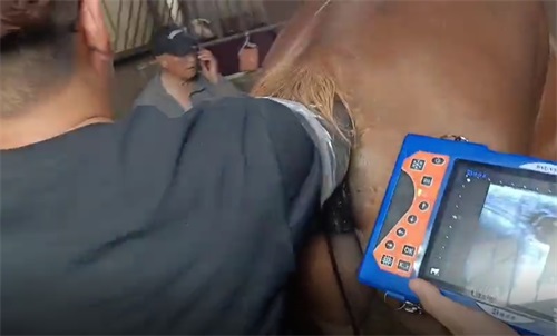
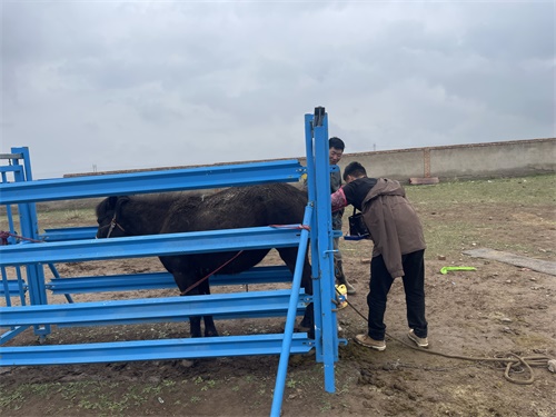
Why Portability Matters
Not all ultrasounds are created equal. For open farm setups, mobility is key. Being able to walk into a pasture or simple handling yard with a lightweight, battery-powered scanner changes everything.
Farmers don't have to move animals long distances or schedule special sessions. The BXL-V50, for instance, is a popular choice because of its long battery life, IP56 waterproof protection, and ease of use in remote or dusty farm environments. With clear B-mode imaging, it’s made for detecting uterine tone, fluid, and even small cysts without needing a power outlet or a controlled clinic environment.
In countries like Canada, where cattle herds are managed across wide landscapes, vets have praised this kind of flexibility. It helps integrate diagnostics into the natural flow of daily farm routines.
Reproductive Timing and Uterine Structure
One major advantage of ultrasound is its ability to monitor uterine involution and readiness for breeding. After calving, the uterus must shrink and clear residual fluids before a cow is fertile again. While this usually happens within 30-45 days, many subfertile cows experience delays.
Ultrasound reveals:
Incomplete involution: The uterus remains large or fluid-filled weeks after calving.
Delayed ovarian rebound: No follicular activity visible in the ovary, which often correlates with uterine issues.
Chronic inflammation: Thickened uterine lining or persistent echogenic material.
Knowing this helps the farmer delay insemination or treat with appropriate antibiotics or hormone protocols, rather than wasting valuable time and semen on a cow that isn’t physiologically ready.
Seasonal Insights and Environmental Influence
Open farms in tropical or arid regions face additional challenges—heat stress and nutrient fluctuations can directly impact uterine recovery and structure. A study conducted across Kenyan smallholder farms showed that cows calving during hot dry seasons had a significantly higher incidence of retained placenta and uterine infections.
With ultrasound, vets could track which cows were bouncing back naturally, and which needed extra intervention. Over time, these farms adjusted their calving plans to avoid peak heat months—something only possible because they had real-time reproductive feedback from portable imaging.
Limitations and Learning Curves
Of course, no tool is perfect. Ultrasound requires training—not just in operating the device, but in interpreting what you see. Two vets can scan the same uterus and reach different conclusions if one isn’t familiar with subtle echotextural signs of pathology.
That’s why building a database of images and cases over time helps. With each scan, you learn what normal looks like on your farm, and how subfertility manifests in your cows.
Ultrasound as a Preventative Tool
More than just a diagnostic for current problems, ultrasound can prevent future issues:
Pre-breeding checks: Confirming uterine tone and absence of fluid before insemination increases conception success.
Postpartum follow-ups: Detecting complications early improves treatment response.
Cull decisions: Identifying chronic uterine issues helps avoid wasting feed and time on cows unlikely to conceive.
On some UK dairy farms, routine ultrasound checks at 21 and 35 days post-calving have become standard practice. The data collected not only improves conception rates but helps predict herd fertility trends for future seasons.
Final Thoughts
Portable ultrasound is no longer a luxury—it’s becoming a core management tool for any farm serious about reproductive efficiency. Especially in open systems where cows breed under less controlled conditions, tracking uterine structure can explain a lot about subfertility that used to remain a mystery.
From catching post-calving complications early to understanding why certain cows keep coming back open, the benefits are clear. It’s about giving cows the best possible chance to conceive—and giving farmers the data they need to make smart, profitable choices.