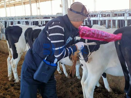
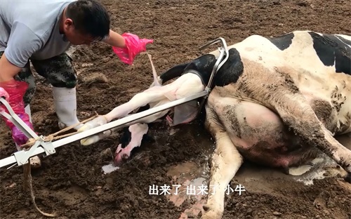
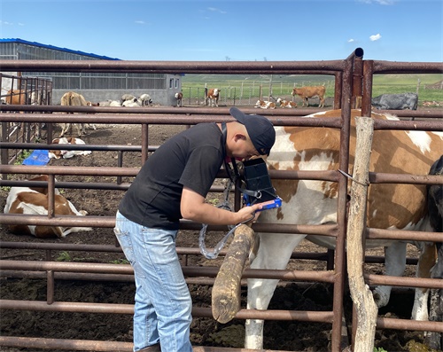
As a wildlife conservation biologist, understanding the reproductive cycles and fetal development in endangered species is essential for effective population management and breeding programs. Among the various monitoring tools available, ultrasonic imaging has become one of the most reliable, non-invasive, and informative methods for evaluating pregnancy status, fetal health, and gestational age in wild mammals. In this article, I’ll share how ultrasound tools are used in reproductive research with endangered wildlife and explore why this technique has become an indispensable element in modern conservation efforts.

Ultrasound in Wildlife Reproductive Studies
Understanding the Importance of Reproductive Monitoring
Many endangered species—from big cats and elephants to primates, marine mammals, and even small marsupials—have low reproductive rates. A single pregnancy can be crucial for a small population, meaning that knowing exactly when an animal is bred, how the embryo is developing, and if the pregnancy is progressing normally can be vital for conservation outcomes.
In captive breeding programs, veterinary teams need to know:
Pregnancy confirmation: Has breeding resulted in conception?
Gestational age: How far along is the pregnancy?
Fetal health: Is the fetus alive and developing normally?
Number of offspring: Will there be twins, triplets, or a singleton birth?
Ultrasonic imaging—especially modern real‑time ultrasonic systems—is one of the few reliable, non‑invasive tools that can answer all of these questions during routine health checks, without exposing the animal to anesthesia or stress repeatedly.
Ultrasonic Principles and Pregnancy Monitoring
What Makes Ultrasonic Imaging Suitable for Wildlife?
Existing across many animal research fields, ultrasonic imaging works by sending high-frequency sound waves into body tissues. The sound echoes are translated into real-time images of internal structures. Key reasons why ultrasonic tools are especially suitable for wildlife reproductive studies include:
Non-invasive: Remote ultrasound probes (transabdominal or transrectal) require only brief physical handling, reducing stress and risk.
Real-time imaging: Pregnancy status, fetal heartbeat, placental location, and even fetal movement can be seen during the scan.
Portable and adaptable: Many ultrasonic units are battery-powered and compact—designed to operate in zoos, sanctuaries, field stations, and even remote research sites.
Versatile frequency use: Depending on the target species, veterinarians can choose lower frequencies for deeper penetration (e.g., large mammals) or higher frequencies for detailed views in small or delicate species.
Monitoring Pregnancy in Different Wildlife Taxa
1. Primates (Great Apes, Monkeys)
Primates—especially great apes—have long gestation periods and complex maternal behaviors. Ultrasonic imaging plays pivotal roles in:
Confirming early pregnancy: In chimpanzees or orangutans, implants can be visualized 30–40 days post-conception.
Measuring fetal size: Crown‑rump length and abdominal circumference help estimate gestational age, which is vital for care planning around birth.
Detecting congenital issues: Organ development (heart, kidneys) can be examined to identify abnormalities early.
2. Carnivores (Tigers, Wolves, Bears)
Large carnivores like tigers and bears present challenges due to thick abdominal muscles and fur. Yet ultrasound is key to:
Breeding program success: Determining pregnancy success early in protected areas and zoos.
Monitoring health: Evaluating fetal well-being—heartbeat, growth, and positioning.
Counting fetuses: Determining litter size informs care planning and helps manage birthing facilities.
3. Elephants and Herbivores
Elephants have one of the longest gestation periods (around 22 months). Ultrasonic monitoring helps:
Track placental development: Since elephants can’t be examined easily, ultrasound probes are sometimes used rectally to follow pregnancy progression.
Estimate gestational month: Fetal measurements support planning for transport, veterinary surveillance, or maternal care.
Smaller herbivores like bison, antelope, or wild goats are frequently scanned ultrasonically to confirm pregnancies and time births to coincide with favorable environmental conditions.
4. Marine Mammals
Cetaceans—dolphins and whales—face additional logistical issues in ultrasound imaging. In aquariums or field environments:
Portable waterproof probes allow visualizing fetal position and health.
Reproductive cycle understanding informs both conservation and research on wild populations.
Pregnancy Stages and Ultrasonic Findings
Early Gestation
Maternal assessment: Uterine size and shape help confirm pregnancy versus pseudopregnancy or reproductive tract abnormalities.
Fetal vesicle detection: A small fluid-filled vesicle may be seen early in the uterus—e.g., ~25–30 days in elephants, similar to domestic cattle—but species-dependent.
Mid-Gestation
Fetal development visibility: Organs, skeletal structures, and heart rate become measurable.
Placental evaluation: Placental adequacy and blood flow—observed in real-time—can preempt complications.
Late Gestation
Fetal positioning and size: Measurements like biparietal diameter indicate fetal growth sufficiency.
Heartbeat and movement: Vital for confirming activity and wellbeing, especially before transport or high-risk births.
Practical Benefits on Conservation Programs
1. Non-Invasive and Low-Risk
Being able to monitor pregnancy without surgery or extended anesthesia increases animal safety. Especially for endangered species, minimizing intervention is critical.
2. Real-Time, High-Value Data
Data on fetal heartbeat, growth, and viability allows timely decisions—whether to transfer a pregnant female, adjust nutrition, or prepare for a high‑risk birth.
3. Resource Optimization
Tracking pregnancy progression helps allocate veterinary and facility resources effectively. For example, managing postpartum care for twins or singletons.
4. Breeding Strategy Enhancement
Data from ultrasound can be correlated with hormone tests or breeding behavior, optimizing timing and success of future matings.
5. Wider Applicability
Ultrasonic protocols developed for one species are often adapted for close relatives or similar-sized animals. For instance, protocols for domestic cattle or horses help shape endangered bovine or equine programs.
Common Ultrasonic Parameters Measured
Much like in cattle growth studies, researchers in wildlife measure several key ultrasonic parameters during pregnancy:
Gestational sac diameter or crown‑rump length
→ Used to estimate age in early pregnancy. Provides a reference curve for each species.Fetal organ sizes and soft‐tissue measurements
→ Includes abdominal circumference, limb length, and thoracic dimensions.Placental thickness and blood flow
→ Detects placental insufficiencies or other threats to viability.Fetal heart rate
→ Vital metric for fetal stress or health; species‑specific values are established from captive populations.Fetal movement observation
→ Normal movement patterns suggest healthy neurological and muscular activity.
Case Study Summaries
Tiger Conservation in Southeast Asia
A conservation team used handheld, battery‑powered ultrasound to confirm pregnancies in several tigers transferred from rescue to a breeding center. By detecting heartbeat and counting four fetuses, they planned nutritional needs and required infrastructure, avoiding overcrowded postpartum areas.
Orangutan Breeding in Zoos
Ultrasonic imaging at 120 days post‐conception allowed veterinary staff to monitor fetal head size and fetal development. Minor abnormalities in limb growth were detected, enabling proactive veterinary elective procedures rather than emergency situations.
Elephant Research in Field Reserves
Using portable trans‑rectal probes, scientists mapped the third‑trimester growth of elephant fetuses in a sanctuarial setting. These measurements helped estimate birth windows, allowing teams to observe natural births and collect critical behavioral data.
Techniques and Equipment
Types of Transducers
Linear array: High‑frequency (7–12 MHz) for shallow imaging in small cats or monkeys.
Curvilinear: Lower frequency (2–5 MHz) for larger mammals like elephants, rhinos.
Intrarectal or intravaginal probes: Used for deeper or less accessible pelvic organs.
Portable Diagnostic Units
Wildlife endocrinology centers and field stations often rely on rugged, battery‑operated ultrasonic units, many with waterproof covers and washable surfaces. Powered ultrasound tools allow routine reproductive monitoring without relying on mains electricity.
Image Analysis Software
Real-time imaging data are recorded and analyzed—often frame by frame—to measure fetal structures, placental dimensions, and blood flow velocity (via Doppler). Digital records support ongoing population health management.
Integrating Data Strategically
Ultrasound findings are often paired with:
Hormone assays (e.g., progesterone, estrogen, relaxin) to refine gestational age estimates and monitor pregnancy health.
Behavioral observations of nests, signs of nesting, or mild isolation before birth in primates and some carnivores.
Genetic tests when verifying lineage in managed breeding programs.
Together, these data enable:
Data-driven decisions like confirming natural breeding success or deciding if artificial insemination is needed.
Managed transfers of pregnant females to areas with better neonatal support.
Long-term record-building for fetal growth norms in endangered species to help future monitoring.
Challenges and Considerations
Species Variation
Protocols vary—gestational sac or fetal size norms differ between species. For lesser-studied mammals, researchers often adapt protocols from domestic livestock or closely related species and validate the data across populations.
Skill Requirements
Achieving consistent, accurate scans demands well-trained technicians or veterinarians. Wildlife clinics now commonly certify staff in point‑of‑care ultrasound, which is becoming a standard skill.
Stress and Restraining Needs
Even though non-invasive, many wildlife ultrasound exams require minimal physical restraint or chemical sedation. Stress management protocols, handling training, and low‑impact techniques help reduce risk during scans.
Equipment Cost and Durability
Advanced ultrasound systems are often expensive and may need regular servicing. For remote operations, ruggedized, portable units with solar or battery charging become essential.
Future Directions in Wildlife Ultrasonography
3D and 4D Imaging
Emerging 3D/4D ultrasonic scanners—capable of reconstructing detailed fetal anatomy—provide richer analysis of fetal health and anomalies, especially for limbs and facial structures.
Artificial Intelligence and Image Recognition
AI systems are under development to assist technicians—automatically measuring structures or detecting deviations from typical growth trajectories—leading to faster, standardized assessments.
Remote Tele-Ultrasound
Advanced units with internet connectivity enable remote veterinarians to assist field teams in validating scans live, reducing training needs and ensuring high-quality data collection.
Multispecies Protocol Standardization
Conservation bodies are working on standardized guidelines for ultrasonic fetal assessment in endangered taxa, improving data comparability across zoos, field programs, and countries.
Why Ultrasonic Tools Have Become Essential
Safety and welfare: No need for surgery; minimal impact on mother and fetus.
Earlier detection: Pregnancy can be confirmed weeks before behavioral or hormonal signs.
Precise timing: Helps schedule births in controlled environments if required or ensures veterinary support.
Enhanced conservation outcomes: Informed breeding improves calf survival; early detection of problems avoids late‑stage pregnancy losses.
Value for money: Despite initial costs, ultrasonic tools help optimize resource use in breeding programs—food, time, personnel.
Building knowledge: Over time, amassed data reveals reproductive norms, typical fetal growth curves, and species-specific health indicators—key for managing small populations.
Conclusion
For wildlife conservationists, reproductive success is at the heart of every endangered‑species recovery plan. Ultrasonic imaging offers a window into the hidden world of fetal development—prompting informed decisions, improving calf survival, and safeguarding the next generation of vulnerable species.
By blending field‑ready hardware, trained veterinary teams, and precise reproductive data, ultrasonic pregnancy tools empower conservation programs to manage breeding with confidence. As protocols improve, AI joins the fray, and imaging technology becomes more accessible, applications will expand—offering a transformative edge in the fight to save endangered wildlife.
References
Esson, M. et al. (2018). Application of ultrasonography in pregnancy management of endangered nonhuman primates. Journal of Zoo and Wildlife Medicine. https://doi.org/10.1638/2017-0177R1
Schmitt, D. et al. (2020). Ultrasound monitoring of fetal development in black rhinos. Conservation Physiology. https://doi.org/10.1093/conphys/coaa020
González, F., & Velázquez, L. (2022). Portable ultrasound tools in aquatic mammal reproductive monitoring. Marine Mammal Science. https://doi.org/10.1111/mms.12982
Wildlife Ultrasound Training Manual. (2024). Wildlife Health & Philanthropy Initiative. https://www.wildlifehealth.org/ultrasound_manual
Smith, A. (2023). Building growth curves: ultrasonographic assessment in endangered ungulates. International Wildlife Symposium Proceedings. https://www.iwsymposium.org/proceedings2023


