Ultrasound imaging is an indispensable tool in equine veterinary medicine, especially when diagnosing tendon injuries in horses. Whether you're managing a performance horse recovering from an injury or conducting routine evaluations for lameness, mastering the technique of tendon ultrasonography ensures accurate assessment and improved rehabilitation outcomes. This guide walks through every step of performing an ultrasound on equine tendons, including preparation, scanning technique, interpretation, and common pitfalls to avoid.

1. Why Ultrasound is Critical for Equine Tendon Evaluation
Tendons in horses—especially the superficial digital flexor tendon (SDFT), deep digital flexor tendon (DDFT), and suspensory ligament—are highly susceptible to strain injuries, particularly in athletic horses. Ultrasound allows for the visualization of tendon architecture, identification of core lesions, fiber alignment, and evaluation of healing over time. Compared to other imaging modalities, ultrasound is non-invasive, relatively affordable, and provides real-time feedback.
2. Equipment Requirements
To perform high-quality tendon ultrasonography, ensure you have the following:
portable ultrasound machine with a linear transducer
Frequency range: 7.5 to 12 MHz (higher frequency provides better resolution but less depth)
Coupling gel (alcohol-based for equine use)
Clippers (if necessary for dense hair coats)
Clean towels and antiseptic scrub

3. Preparing the Horse and the Area
Preparation is key to obtaining clear images:
Restrain the horse in a calm, well-lit environment. Cross-ties or stocks are recommended.
Identify the limb and tendon to be scanned (typically the palmar aspect of the metacarpal region for front limbs).
Clip the hair from the scanning area to improve contact and reduce air artifacts.
Clean the area with a mild antiseptic solution to remove dirt, sweat, or oils.
Apply ultrasound gel liberally to ensure good acoustic coupling.
4. Scanning Technique
A systematic scanning protocol improves consistency and allows for comparison over time or between limbs.
a. Limb Position: Ensure the limb is weight-bearing and perpendicular to the ground. Tendons are most relaxed and easier to image in a neutral standing position.
b. Imaging Planes:
Longitudinal (sagittal) plane: Probe is aligned parallel to the tendon fibers.
Transverse (axial) plane: Probe is rotated 90° to capture cross-sectional views.
c. Scanning Zones: Divide the metacarpal or metatarsal region into six zones (Zone 1A to 3B), each 4 cm in length, starting from the distal aspect of the accessory carpal bone. Scan each zone in both longitudinal and transverse planes.
d. Image Acquisition: Adjust gain, depth, and focus to optimize the image. Tendons should appear as a linear, fibrillar pattern in longitudinal views and a round to oval, echogenic structure in transverse views. Identify any areas of hypoechogenicity (dark regions), fiber disruption, or swelling.

5. Interpreting the Images
Tendon lesions typically appear as areas of decreased echogenicity (dark areas) with disrupted fiber alignment. Important diagnostic parameters include:
Lesion size (measured in both planes)
Percentage of cross-sectional area affected
Echogenicity (anechoic, hypoechoic, or mixed)
Fiber pattern (linear, disrupted, or chaotic)
Presence of tendon sheath effusion
Compare the injured limb with the contralateral (unaffected) limb whenever possible. This helps identify subtle changes and provides a baseline for follow-up exams.
6. Common Injuries Identified by Ultrasound
Core Lesions: Central hypoechoic regions, common in SDFT injuries
Marginal Tears: Disruption at the edges of the tendon
Suspensory Ligament Desmitis: Diffuse swelling and decreased echogenicity
Tendon Sheath Effusion: Fluid accumulation around the tendon indicating inflammation

7. Follow-Up and Monitoring
Ultrasound is invaluable for monitoring tendon healing. Repeat scans are typically conducted every 30 to 60 days during rehabilitation. Progressive improvements should show increased echogenicity and restoration of fiber alignment. Scanning should continue until tendon architecture returns to near-normal and training can be resumed gradually.
8. Tips for Better Imaging and Fewer Errors
Always scan both limbs for comparison.
Maintain consistent limb positioning and scanning technique across sessions.
Use ample gel and avoid air bubbles between the probe and skin.
Start with lower gain and adjust upward to avoid overexposure.
Record and archive all images with zone labels and date for future reference.
9. Limitations and Complementary Diagnostics
While ultrasound is excellent for soft tissue evaluation, it has limitations. It does not visualize bone or deep ligamentous structures well. In complex cases, MRI or CT may be required. Clinical assessment, lameness exams, and nerve blocks should always accompany ultrasound findings for a comprehensive diagnosis.

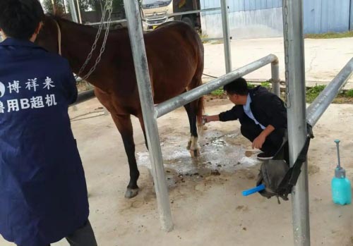
BXL-DZ20 Horse Tendon Ultrasound Instrument
10. Working with Your Veterinarian
If you're a farm manager, breeder, or trainer performing routine scans, always consult with an equine veterinarian for initial diagnosis and interpretation. Many lesions can look similar on ultrasound, and accurate reading requires experience and a trained eye. Vets can also provide a tailored rehabilitation protocol based on lesion type and severity.
Conclusion
Equine tendon ultrasonography is an essential skill in modern horse care. When performed correctly, it provides accurate and actionable information on soft tissue injuries, helping prevent further damage and guiding effective treatment. By understanding the anatomy, mastering the scanning technique, and interpreting the images reliably, you can play a vital role in the health, performance, and longevity of your horses. Whether you're a veterinarian, breeder, or dedicated caretaker, integrating ultrasound into your routine care will elevate your standard of equine management.