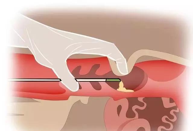
When using a B-ultrasound probe to detect cows, the bladder is displayed in sequence, followed by the cervix and uterine body, and then the uterine angle. The probe is kept horizontal and located in the median plane of the pelvic canal. You can also start from the skull part of the horn and then follow the path of the two horns towards the vagina in sequence
")
Cattle use B-ultrasound probe to detect position
The ultrasound appearance of a cow's uterus changes according to the time and stage of the cycle (follicular phase/luteal phase) after calving.
In the early postpartum period, the flesh nodes are still visible and there is still a large amount of fluid in the uterine cavity with echoes (only slightly lower than the uterine wall). Wall thickness, blood vessels are clearly visible on its thickness (circular or elliptical anechoic zone). Gradually, the amount of liquid will decrease and the flesh will no longer be visible. 21 days postpartum, uterine involution is completed, and the ultrasound image is similar to that of the luteal phase uterus.
")
Cross section of postpartum uterus in cows
Cross section of postpartum uterus. The uterine wall is circled in blue. There is fluid accumulation (green) in the uterine cavity. There is also an ovary circled in black and a thorn (highlighted in red)