The animal mammary gland is located in a relatively shallow part, so the high-frequency (7.5-14MHz) veterinary B-ultrasound probe with weak penetration should be selected to enable a clearer display of the internal structure of the mammary gland. The ultrasonography of normal animal mammary gland consists of four layers, skin, subcutaneous fat, gland and post-mammary structure, from shallow to deep. The skin is a strong echo zone, about 2mm thick, regular and smooth outline, thicker in the areola and lower quadrant; In animal B-ultrasound, animal papillae are round or oval with medium or strong echo, and the deep boundary is usually smooth. Decreased echo or sound attenuation is common under the nipple areola, mainly due to the more fibrous tissue in this part and the tendency of the catheter to vertical. The subcutaneous fat layer is a low-echo lobular structure with unclear boundaries. The glandular layer (also called parenchymal layer) echo pattern varies with age and is mainly composed of glandular tissue, including the breast lobules and the lactation duct system, and the fibrous connective tissue between them. In animal B-ultrasound, the former is low echo, the latter is strong echo, and the different ratio or composition of the two determines the echo intensity of this layer or some areas in the layer. Relatively speaking, there are more glandular tissues with low echo in this layer, which decrease slowly after entering the later stage, while the decrease rate of fibrous connective tissue is slower, so the strong echo component increases. In animal B-ultrasound, there is a strong echo between the glandular layer and the skin of the animal suspensory ligament (Cooper ligament), which is similar to triangle or contact, and continues in the fibrous connective tissue within the glandular layer.

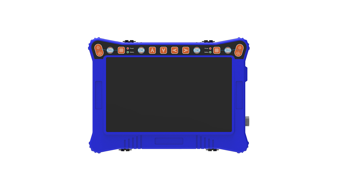
Veterinary B-ultrasound BXL-V30
Note on images ① Read veterinary B-ultrasound images, and do not observe an indicator of ultrasound alone (such as no echo is not necessarily a benign cyst). (2) Comprehensive ultrasonic indicators such as lesion size, shape, boundary, echo, internal structure, rear echo and the relationship with surrounding tissue should be comprehensively observed for analysis. (3) Veterinary B ultrasound can be continuously observed, compared before and after images, and combined with clinical symptoms and laboratory results for comprehensive analysis.


