For horse owners, nothing sparks more immediate concern than the onset of colic. It’s one of the most common emergencies in equine practice, and it ranges from mild gas buildup to severe intestinal twists that can be fatal. Over the years, farmers and veterinarians have refined their skills in identifying early signs of colic, but now, with the help of modern ultrasound scanning, it’s possible to get a clearer, more accurate look inside a horse’s abdomen—often before symptoms become critical.
")
Let’s talk honestly: colic is scary. Horses are stoic animals, and many don't show outward signs until the pain becomes severe. Even experienced farmhands can sometimes miss the early indicators. But when ultrasound technology comes into play, it gives everyone—from stable managers to rural vets—a real-time view of what’s happening inside.
Why Farms Are Turning to Ultrasound for Colic Monitoring
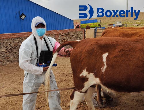
Historically, diagnosing colic on a farm relied mostly on physical exam, gut sounds, and rectal palpation. These are useful tools, but they don’t always provide a full picture. Ultrasound, on the other hand, allows the vet (or in some cases, trained farm staff) to non-invasively scan the horse's abdomen, visualize loops of intestine, detect fluid buildup, and monitor motility in real time.
What’s changed in recent years is that portable ultrasound machines have become more affordable and user-friendly. Instead of relying on large referral hospitals, farms now have access to veterinary-grade scanners that can be used right in the stall. This shift is empowering farms to act faster and with more confidence.
Recognizing the First Signs of Colic
Every horse is different, but common early signs of colic include:
Pawing at the ground
Looking at or kicking the belly
Rolling or lying down repeatedly
Restlessness or refusing to eat
Abnormal gut sounds (or lack of them)
These signs can escalate quickly. That’s where ultrasound scanning steps in—not to replace traditional exams, but to strengthen and confirm clinical impressions. Within minutes of applying the probe, it’s often possible to see whether the intestines are distended, filled with gas, or not moving as they should.
What Exactly Can Ultrasound Show in a Colicky Horse?
There are several key markers that ultrasound can detect when colic is suspected:
Intestinal distension: If bowel loops appear larger than normal, it suggests blockage or gas accumulation.
Free abdominal fluid: More than a few milliliters could mean something serious, like bowel rupture or inflammation.
Reduced motility: Healthy intestines should be visibly moving. A "silent" gut on ultrasound raises red flags.
Thickened intestinal walls: This could indicate inflammation, infection, or compromised blood flow.
Farmers working with a trusted vet can use ultrasound to track changes over time. For example, is the gut filling with more fluid, or is it starting to empty? Is motility returning after pain relief or hydration? These insights help make decisions about whether to continue treatment at the farm or prepare for referral to an equine hospital.
Real Stories from the Field
Many farmers across Europe and North America are now integrating ultrasound into their emergency toolkits. Take the case of a Welsh pony farm in rural England: one young mare began showing vague signs of colic—mild pawing and a slight loss of appetite. The farm manager, trained to use a portable ultrasound under vet supervision, scanned her abdomen. The scan revealed gas distension but no signs of fluid or twisted loops. With this reassurance, the mare was given anti-spasmodic treatment, closely monitored, and recovered within hours—avoiding a stressful (and expensive) clinic visit.
Or consider a Texas ranch dealing with chronic colic in a retired barrel racing gelding. By monitoring gut motility weekly via ultrasound, they identified patterns related to feed changes and seasonal stress. Adjustments to his hay and turnout routine drastically reduced his colic episodes.
These real-life examples show how practical ultrasound can be—not just for emergencies, but also for long-term gut health management.
Advantages of Using Ultrasound on the Farm
Non-invasive and painless: Horses don’t need to be sedated. The probe is simply placed on the skin with gel, often while the horse is standing.
Immediate insights: In urgent situations, time matters. Ultrasound provides real-time data without needing to wait for bloodwork or other diagnostics.
Fewer unnecessary referrals: Not every colic needs surgery or hospitalization. Ultrasound helps distinguish mild from serious cases.
Cost-effective in the long run: One bad colic case can cost thousands. Early diagnosis with ultrasound can prevent complications, saving both lives and money.
Is It Complicated to Use?
For licensed veterinarians, Equine Ultrasound is already part of their diagnostic toolkit. But more and more farms are getting trained in basic scanning under a vet’s guidance. The idea isn’t to replace the vet—it’s to gather better info faster and communicate clearly when a vet is called in.
Handheld machines are designed for rugged farm use. Most come with equine presets and intuitive interfaces. A few practice sessions are often enough for farm managers to feel comfortable identifying the cecum, small intestine, or stomach area—and when something just doesn’t look right.
Ultrasound vs. Other Diagnostic Tools
Rectal palpation can detect some types of impaction or displacement but carries a small risk of rectal tears, especially with fractious horses.
Blood tests like lactate levels are useful but don’t give structural information.
Nasogastric tubing helps relieve pressure but doesn’t tell you why there’s pressure to begin with.
X-rays are limited in abdominal imaging due to the horse’s size.
Ultrasound, on the other hand, is fast, safe, and highly visual. It complements other diagnostic tools rather than replacing them.
A Look at Colic Prevention with Ultrasound
Interestingly, ultrasound isn’t just for emergencies anymore. Farms are beginning to use it as a preventive tool, especially for horses with a history of colic. Regular scans can help detect slow motility, intestinal wall thickening, or gas buildup before symptoms even appear.
Think of it like a window into the gut—a chance to catch subtle changes and adapt feeding, turnout, or hydration plans accordingly. Prevention is always better than treatment, especially with something as unpredictable as colic.
Final Thoughts from the Barn Aisle
At the end of the day, horse health is all about timing. The earlier you can act, the better the outcome. That’s why ultrasound scanning is making such a big difference on farms. It doesn’t take away the need for experience, observation, and veterinary input—but it adds a whole new level of insight.
For farms serious about equine health, having an ultrasound scanner on hand is no longer a luxury—it’s quickly becoming a necessity. It saves time, lowers stress, and, most importantly, improves survival rates when colic strikes.