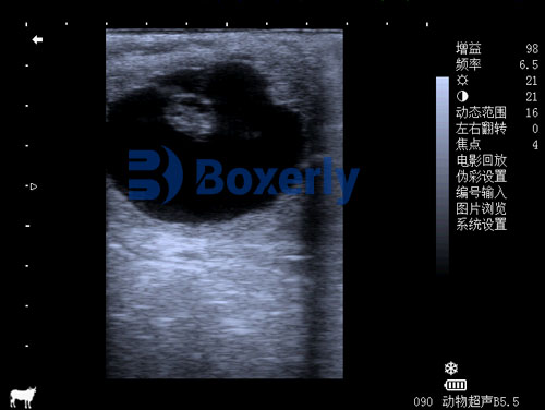
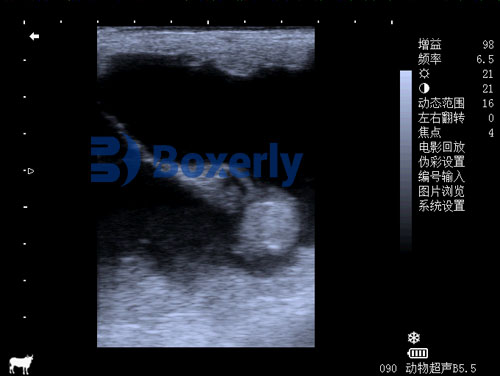
For breeders, veterinarians, and equine managers, the early weeks of a mare’s pregnancy are critical. Identifying whether the pregnancy is viable, healthy, and progressing normally can save time, money, and improve the welfare of both mare and foal. In recent years, ultrasound technology has emerged as the go-to diagnostic tool for equine reproductive assessment—especially during early gestation. From confirming pregnancy to detecting uterine abnormalities and fetal viability, ultrasound (sonography) provides real-time, non-invasive insight into equine maternal health.

In this article, I’ll explore the key ultrasound indicators of health in pregnant horses during early gestation, combining both veterinary best practices and real-world knowledge from international sources and studies. Whether you’re a professional veterinarian or a horse owner involved in breeding, this guide will help you understand what to look for on an ultrasound and how to interpret the findings in the early stages of a mare’s pregnancy.
The Importance of Early Gestation Monitoring in Horses
In horses, early gestation typically refers to the first 30–45 days post-ovulation. During this period, the embryo undergoes rapid development, while the mare’s uterus adapts to support the growing fetus. However, this is also the most vulnerable phase—up to 20% of equine pregnancies fail during this time, often without external signs.
This is why equine practitioners globally recommend early ultrasound checks at day 14, day 25, and day 35 after ovulation. These checks not only confirm pregnancy but also provide critical health indicators that guide management decisions.
Key Ultrasound Health Indicators in Pregnant Mares
1. Embryonic Vesicle Detection (Day 11–16)
The first reliable sign of pregnancy is the embryonic vesicle, a fluid-filled spherical structure that can be visualized as early as day 11 post-ovulation using transrectal ultrasonography. By day 14, this vesicle should measure 9–15 mm in diameter and appear as a clear, round structure in the uterine lumen.
Indicator of health: A well-defined, symmetrical vesicle suggests normal embryonic development. Irregular shapes or poor mobility may indicate developmental delays or uterine fixation problems.
2. Fixation of the Vesicle (Day 16)
By day 16, the vesicle usually “fixes” in one spot in the uterus—most commonly at the base of the uterine horn. This process is necessary for successful placental development.
Indicator of health: Lack of fixation by day 17 or abnormal location (e.g., near the cervix) may signal a risk of pregnancy loss or complications.
3. Detection of the Embryo Proper and Heartbeat (Day 21–28)
Between days 21–25, the embryo proper becomes visible as a small structure inside the vesicle. By day 25, a flickering heartbeat can often be seen using B-mode ultrasound, confirming a viable pregnancy.
Indicator of health: Presence of heartbeat is the most reliable sign of fetal viability. A non-pulsating embryo or reduced heart rate may indicate stress or early embryonic death.
4. Yolk Sac and Allantois Development (Day 25–35)
As pregnancy progresses, two important fluid compartments—the yolk sac and the allantois—become distinguishable. A balanced growth of these structures ensures proper nutrient exchange and waste removal.
Indicator of health: Disproportionate fluid compartments or collapsed yolk sacs may point to developmental anomalies.
5. Uterine and Cervical Tone
Even in early pregnancy, ultrasound can assess the tone of the uterus and cervix. A firm, toned uterus and tightly closed cervix suggest a healthy environment for the embryo.
Indicator of health: Soft or fluid-filled uterus, or open cervix, may indicate uterine infection (endometritis), twin pregnancy risks, or hormonal imbalances.

Common Pathologies Detected by Ultrasound in Early Gestation
Ultrasound is not only about confirming pregnancy—it’s essential in detecting abnormalities that can lead to early pregnancy failure. Here are some of the most common findings during early checks:
1. Twin Pregnancies
While rare in some countries, twin pregnancies occur in 10–15% of thoroughbred mares, and over 90% result in pregnancy failure if not managed. Ultrasound at day 14 is critical for identifying twins while they are still mobile and suitable for manual reduction.
2. Anembryonic Pregnancy (Blighted Ovum)
This is characterized by a normal-looking vesicle that never develops an embryo. Ultrasound by day 25–30 will show an empty vesicle with no embryo or heartbeat.
3. Early Embryonic Death (EED)
Signs include a previously visible heartbeat that disappears, or a shrinking and collapsing vesicle. EED occurs in up to 20% of equine pregnancies and is most common before day 35.

Ultrasound Techniques Used in Early Equine Pregnancy
Transrectal B-mode Ultrasound
This is the standard method for early pregnancy diagnosis in mares. It provides real-time black-and-white imaging and allows veterinarians to examine the uterus, ovaries, and embryo in great detail.
Color Doppler Imaging (Optional)
In some high-risk cases, Color Doppler may be used to assess blood flow to the embryo and uterus, helping detect inflammation or inadequate placental circulation.
Using Portable Ultrasound Units
Many veterinarians now use portable ultrasound units in field settings, like the BXL-V50, which offers high-resolution imaging and long battery life—ideal for working on farms without constant electricity. Its waterproof and durable design ensures reliable readings, even in tough rural conditions.

International Perspectives on Equine Pregnancy Ultrasound
Across the world, veterinarians share common goals: improve foal survival, reduce costs, and ensure mare welfare. In North America and Europe, ultrasound is standard for thoroughbred and sport horse breeding programs. Countries like Germany, Australia, and the UK enforce tight breeding schedules and rely on precise ultrasound data to optimize mating and embryo transfer.
In South America and Asia, portable ultrasound machines have made early diagnosis more accessible to mid-sized breeders and veterinarians in rural areas. Studies from Brazil and Argentina highlight how early embryonic detection has reduced loss rates and improved breeding timelines.
Veterinary training programs worldwide now include detailed instruction on early gestational scanning, and published research continues to refine our understanding of embryonic development stages.

Practical Benefits for Breeders and Horse Owners
Using ultrasound during early gestation allows for:
Precise breeding management: Re-checking mares ensures timely re-breeding if a pregnancy fails.
Economic efficiency: Avoid wasting resources on non-pregnant mares.
Improved foal survival: Early detection of abnormalities allows for timely intervention.
Better record-keeping: Visual confirmation and documentation aid in reproductive performance tracking.
Summary of Key Ultrasound Indicators in Early Pregnant Mares
| Parameter | Normal Findings (Day 11–35) | Abnormal Findings |
|---|---|---|
| Embryonic Vesicle | Round, mobile, 9–30 mm | Misshapen, non-mobile |
| Fixation | Occurs by Day 16 at horn base | Delayed fixation or cervical fixation |
| Embryo Proper | Visible by Day 21–25 | Absent or malformed |
| Heartbeat | Detected by Day 25 | Absent or bradycardic |
| Fluid Compartments | Balanced yolk sac and allantois | Collapsed yolk sac |
| Uterine/Cervical Tone | Firm uterus, closed cervix | Flaccid uterus, open cervix |
| Twin Pregnancy | Two vesicles in separate horns (early detection vital) | Overlapping vesicles or late detection |
Conclusion
Ultrasound technology is transforming the way equine pregnancies are managed, especially during the vulnerable early stages. By providing real-time insights into embryonic development, uterine health, and fetal viability, it enables timely and informed decisions that can improve breeding outcomes and save lives.
From confirming the presence of a viable embryo to detecting high-risk conditions like twin pregnancies or early embryonic death, ultrasound has become the most effective tool in early gestation monitoring for horses. Its adoption by breeders and veterinarians worldwide speaks to its value, reliability, and impact.
As more portable and high-resolution devices like the BXL-V50 enter the market, even remote or mid-scale farms can gain access to advanced reproductive diagnostics—making equine breeding safer, smarter, and more efficient.
References:
McCue, P. M. (2014). Ultrasound Evaluation of Early Pregnancy in the Mare. Colorado State University Equine Reproduction Laboratory. https://csu-cvmbs.colostate.edu/vth/Pages/equine-reproduction.aspx
Ginther, O. J. (1995). Ultrasonic Imaging and Animal Reproduction: Horses. Equiservices Publishing.
Youngquist, R. S., & Threlfall, W. R. (2007). Current Therapy in Large Animal Theriogenology. Elsevier Health Sciences.
Rantanen, N. W. (1993). Use of Diagnostic Ultrasound in the Equine. Veterinary Clinics of North America: Equine Practice.
Squires, E. L., & Carnevale, E. M. (2021). Equine Embryo Transfer and Assisted Reproduction. Frontiers in Veterinary Science. https://www.frontiersin.org/articles/10.3389/fvets.2021.661765/full


