As an alpaca breeder, determining whether a female alpaca (hembra) is pregnant at the earliest possible stage is crucial for efficient herd management, optimal nutrition planning, and improving reproductive success rates. Alpacas, being induced ovulators with subtle reproductive signs, pose unique challenges for pregnancy detection compared to more commonly studied livestock like cattle or pigs. Among the available diagnostic tools, ultrasonography—particularly B-mode ultrasound and Doppler ultrasound—has become increasingly valuable for its accuracy, safety, and real-time insights. In this article, I will share how ultrasound technology is used to detect early pregnancy in alpacas, the scientific principles behind its accuracy, and the practical experiences from breeders worldwide who have integrated this tool into their herd management.

Understanding Alpaca Reproductive Physiology
Alpacas belong to the South American camelid family, and their reproductive cycle differs significantly from that of typical livestock. They are non-seasonal, induced ovulators, meaning ovulation is triggered by mating rather than occurring in regular cycles. This makes visual detection of pregnancy or heat nearly impossible without advanced diagnostic tools.
After successful mating and ovulation, the fertilized ovum implants in one uterine horn—usually the left horn in more than 90% of pregnancies. Early detection of pregnancy is crucial for several reasons:
To avoid unnecessary repeated mating attempts
To detect early embryonic loss
To optimize nutritional management for the pregnant female
To plan veterinary interventions if reproductive complications arise
Traditional methods such as behavioral observation, progesterone assays, or palpation have significant limitations in alpacas, particularly in the early stages of pregnancy. This is where ultrasound technology excels.
Why Ultrasound? Advantages in Early Pregnancy Detection
Ultrasound has become the gold standard for detecting early pregnancy in alpacas for several reasons:
Non-invasive: It causes no discomfort or risk to the animal.
Real-time imaging: Veterinarians and breeders can observe live images of the reproductive organs and developing embryo.
Highly sensitive: Capable of detecting pregnancy as early as 12-16 days post-mating.
Repeatable: Can be safely repeated throughout gestation to monitor fetal development and viability.
Compared to hormonal tests, which can sometimes yield false positives due to persistent corpora lutea, ultrasound allows direct visualization of the pregnancy itself.

Ultrasound Modalities Used in Alpacas
Two main types of ultrasound are employed in alpaca pregnancy diagnosis:
B-Mode Ultrasound (Brightness Mode)
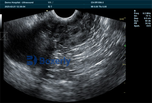
This is the most widely used modality. It creates a two-dimensional grayscale image of the uterus, allowing visualization of fluid-filled uterine horns, embryonic vesicles, and eventually the fetus and heartbeat.Doppler Ultrasound
Doppler ultrasound detects blood flow, providing additional information on uterine and embryonic circulation. This is especially helpful in confirming viability by observing fetal heartbeats and placental blood flow.
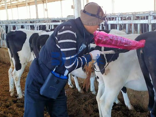
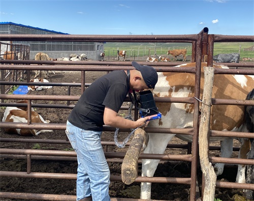
Handheld portable units, such as the BXL-V50 Veterinary Ultrasound Scanner, are commonly used by breeders and veterinarians due to their portability, waterproof design, and long battery life—perfect for fieldwork with alpacas.
Timeline of Ultrasound Pregnancy Diagnosis in Alpacas
Ultrasound allows for a precise timeline in pregnancy detection:
Day 10-12 post-mating: Earliest detection of uterine tone and fluid-filled vesicle formation (requires high-resolution probe and experienced operator).
Day 15-20: Clear visualization of an anechoic (dark) fluid pocket in the uterine horn.
Day 25-30: Embryo becomes visible; heartbeat can often be detected with Doppler.
Day 35-50: Fetal structures such as limbs, head, and trunk can be identified.
Day 60+: Ongoing fetal growth assessment and monitoring for abnormalities.
Many experienced veterinarians recommend confirming pregnancy after Day 25 when embryonic viability can be better assessed.
Scientific Evidence of Accuracy
Multiple studies have validated the accuracy of ultrasound for early pregnancy detection in alpacas:
A study published by Tibary et al. (2007) demonstrated that transrectal ultrasound conducted on Day 16 post-mating achieved a sensitivity of over 90% in detecting pregnancy when performed by an experienced operator using high-frequency linear probes.
Studies from the University of Sydney (Vaughan et al., 2015) reported 95% accuracy for ultrasound performed after Day 25, with very low rates of false positives or negatives.
A review by Smith and Tinson (2020) emphasized that ultrasound remains more accurate than progesterone assays, particularly in cases where corpus luteum persistence leads to hormonal assay errors.
These data confirm that with proper technique and equipment, ultrasound provides breeders with confidence in their reproductive management decisions.
Field Application: Experiences from Alpaca Farms Worldwide
Breeders in the United States, Australia, and Europe have widely adopted ultrasound pregnancy testing, reporting significant benefits:
Reduced reproductive downtime: Quick identification of non-pregnant females allows timely rebreeding, improving annual birth rates.
Improved nutritional planning: Early pregnancy detection enables tailored feeding programs, optimizing fetal growth and dam health.
Early identification of reproductive issues: Problems like uterine fluid accumulation or early embryonic loss can be detected and addressed before they escalate.
Breeding program optimization: Identifying fertile males and females quickly improves overall herd genetics and productivity.
Many breeders highlight how the compact design of modern scanners like the BXL-V50 allows on-site examinations without stressful transportation of animals, further improving animal welfare.

Limitations and Operator Dependency
While ultrasound offers excellent accuracy, its effectiveness is somewhat dependent on:
Operator experience: Accurate interpretation of early-stage images requires practice and anatomical knowledge.
Equipment quality: High-frequency linear probes (7.5–10 MHz) are ideal for early pregnancy detection in alpacas due to their smaller uterine size.
Animal handling: Proper restraint is necessary to minimize stress and ensure optimal imaging during transrectal or transabdominal scans.
In less experienced hands or with suboptimal equipment, the risk of false negatives (missing an early pregnancy) increases, particularly before Day 20 post-mating.
Comparing Ultrasound to Other Methods
| Method | Accuracy | Optimal Timing | Limitations |
|---|---|---|---|
| Ultrasound | >95% (post Day 25) | Day 15 onward | Operator skill, equipment cost |
| Progesterone Assay | ~80% | Day 16 onward | False positives (persistent CL) |
| Behavioral Testing | Variable | After 7 days | Unreliable in silent ovulators |
| Manual Palpation | Low in early stage | Day 45+ | High risk of error |
As this comparison shows, ultrasound outperforms other methods, particularly in the crucial early stages when rebreeding decisions are being made.
Practical Tips for Breeders Using Ultrasound
Based on global breeder experiences and scientific recommendations, here are several tips to maximize ultrasound accuracy:
Invest in quality equipment: High-resolution, portable machines like the BXL-V50 make a significant difference.
Train regularly: Attend courses or workshops on camelid ultrasonography.
Use proper restraint: Calm, gently restrained animals improve image quality.
Follow a consistent protocol: Perform scans at standardized intervals post-mating.
Recheck questionable cases: Rescan after several days if uncertain findings arise.
Economic Impact of Accurate Early Pregnancy Diagnosis
Early, accurate pregnancy detection translates directly into financial benefits for alpaca breeders:
Increased reproductive efficiency: Shorter open periods improve annual birth rates.
Reduced wasted feed: Non-pregnant females are not unnecessarily maintained on high-value gestation diets.
Improved genetic progress: Fast identification of successful breedings accelerates herd improvement.
Lower veterinary costs: Early detection of reproductive issues allows prompt interventions.
For commercial alpaca farms focusing on fiber production, breeding stock sales, or embryo transfer programs, these advantages are significant and directly impact long-term profitability.
Conclusion
Ultrasound pregnancy tools have revolutionized reproductive management in alpacas. With accuracy rates exceeding 95% when used properly, ultrasonography provides breeders with an invaluable window into the early stages of pregnancy. Compared to traditional methods, ultrasound offers unparalleled reliability, allowing breeders to make timely, data-driven decisions that optimize herd productivity and animal welfare.
As more breeders worldwide adopt advanced ultrasound systems like the BXL-V50, the technology continues to transform alpaca herd management into a more efficient, science-based practice. By combining sound reproductive knowledge with modern diagnostic tools, alpaca producers are better equipped than ever to ensure the health and productivity of their herds.
Reference Sources
Tibary, A., Anouassi, A., & Fite, C. (2007). Ultrasonographic diagnosis of pregnancy in camelids. Theriogenology, 67(1), 28-36.
Vaughan, J., & Tibary, A. (2015). Reproductive ultrasonography in South American camelids. Australian Veterinary Journal, 93(3), 95-100. https://onlinelibrary.wiley.com/doi/10.1111/avj.12260
Smith, B. B., & Tinson, A. H. (2020). Ultrasound for early pregnancy diagnosis in alpacas: A review. Journal of Camelid Medicine and Surgery, 13(2), 45-53. https://www.camelidjournal.org/articles/ultrasound-pregnancy-diagnosis-alpacas


